


Yolk sac fetal pole no heartbeat: Fetal Pole: Ultrasound, Anatomy & Function
Abnormal Ultrasound in Early Pregnancy
« Back to Articles
Dr. Vishvanath Karande
Oct 28
222 Comments
In a previous blog we discussed the expected ultrasound findings in a normal intrauterine pregnancy. Variations from the expected pattern of development are worrisome or, if major, definitive for early pregnancy failure or miscarriage. These were discussed in a recent review article by Doubilet et al. (N Engl J Med 2013;369:1443-51). Here is a summary:
The criteria most often used to diagnose pregnancy failure are the absence of cardiac activity by the time the embryo has reached a certain length (crown–rump length), the absence of a visible embryo by the time the gestational sac has grown to a certain size (mean sac diameter), and the absence of a visible embryo by a certain point in time.
Crown-rump length (CRL)
A crown–rump length of 5 mm was widely recommended as a positivity criterion for diagnosing failed pregnancy when no cardiac activity is seen. Recent studies have shown that a 5-6 mm cutoff can result in a false positive diagnosis of pregnancy failure. It is now recommended that we use a 7 mm (rather than 5 mm) cut-off for diagnosing failed pregnancy. Thus if the crown-rump length is 7 mm and there is no heart beat visible, it is suspicious for a failed pregnancy.
Gestational sac diameter
It is prudent to use a cutoff of 25 mm (rather than 16 mm) for the mean sac diameter with no visible embryo in diagnosing failed pregnancy (see figure above). This would yield a specificity and positive predictive value of 100% (or as close to 100% as can be determined). When the mean sac diameter is 16 to 24 mm, the lack of an embryo is suspicious for, though not diagnostic of, failed pregnancy
Time based criteria for failed pregnancy
Not all failed pregnancies ever develop a 7-mm embryo or a 25-mm gestational sac, so it is important to have other criteria for diagnosing pregnancy failure. The most useful of such criteria involve non visualization of an embryo by a certain point in time. An alternative approach to predicting pregnancy failure, based on subnormal growth of the gestational sac and embryo, has been shown to be unreliable. Non visualization of an embryo with a heart- beat by 6 weeks after the last menstrual period is suspicious for failed pregnancy, but dating of the last menstrual period (in a pregnancy conceived without medical assistance) is too unreliable for definitive diagnosis of pregnancy failure.
The timing of events in early pregnancy — gestational sac at 5 weeks, yolk sac at 5 ½ weeks, and embryo with heartbeat at 6 weeks — is accurate and reproducible, with a variation of about ± ½ week; this consistency explains the time-related criteria for pregnancy failure. For example, if the initial ultrasonogram shows a gestational sac with a yolk sac and a follow-up scan obtained at least 11 days later does not show an embryo with cardiac activity, the diagnosis of failed pregnancy is established.
According to the Society of Radiologists in Ultrasound Multispecialty Consensus Conference on Early First Trimester Diagnosis of Miscarriage and Exclusion of a Viable Intrauterine Pregnancy, October 2012; the following are guidelines for Transvaginal Ultrasonographic diagnosis of Pregnancy Failure in a Woman with an Intrauterine Pregnancy of Uncertain Viability.
Findings Diagnostic of Pregnancy Failure
- Crown–rump length of ≥7 mm and no heartbeat
- Mean sac diameter of ≥25 mm and no embryo
- Absence of embryo with heartbeat ≥2 wk after a scan that showed a gestational sac without a yolk sac
- Absence of embryo with heartbeat ≥11 days after a scan that showed a gestational sac with a yolk sac
Findings Suspicious for, but Not Diagnostic of, Pregnancy Failure
- Crown–rump length of <7 mm and no heartbeat
- Mean sac diameter of 16–24 mm and no embryo
- Absence of embryo with heartbeat 7–13 days after a scan that showed a gestational sac without a yolk sac
- Absence of embryo with heartbeat 7–10 days after a scan that showed a gestational sac with a yolk sac
- Absence of embryo ≥6 wk after last menstrual period
- Empty amnion (amnion seen adjacent to yolk sac, with no visible embryo)
- Enlarged yolk sac (>7 mm)
- Small gestational sac in relation to the size of the embryo (<5 mm difference between mean sac diameter and crown–rump length)
When there are findings suspicious for pregnancy failure, follow-up ultrasonography at 7 to 10 days to assess the pregnancy for viability is generally appropriate. Treatments for early miscarriage are discussed here.
To see a fertility specialist who is a board-certified physician with high success rates, make an appointment at one of InVia’s four Chicago area fertility clinics.
Is no fetal heartbeat at 6 weeks of pregnancy a sign of miscarriage? –
Is no fetal heartbeat at 6 weeks of pregnancy a sign of miscarriage?
Miscarriage is one of the most stressful situations for a couple who are trying hard for conceiving. Such couples if achieve pregnancy but no fetal heartbeat is visible, can assume the condition as a miscarriage. Is it really so? In this blog, Dr. Nishant Dixit, a highly experienced Reproductive Medicine Specialist will explain how and at what time embryos grow in the uterus and what is delayed conception? Moreover, causes of no heartbeat at 6-week conception are also discussed, and how the couple should proceed in such a situation?
In 15–20% cases out of 100 pregnancies, the fetal heartbeat is not detected within 6 weeks of internal sonography.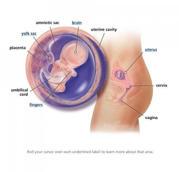 The situation can be very depressing because couples who have conceived but there’s no fetal heartbeat, such couples begin to panic as they generally interpret it as a pregnancy loss. Such couples always think about what can be done to bring the heartbeat of the baby. To understand the process of fetal heartbeat detection, it is important to know the timeline of naturally conceived pregnancy and how different milestones are achieved in the process.
Heartbeat detection in natural pregnancy
For example, if ovulation in women occurs on the fourteenth day, then after 5-7 days, the embryo begins to develop in the fallopian tube and thereafter it rolls down to the uterus and is implanted in the uterus wall. As soon as the embryo is implanted, the embryo’s cells begin to divide and secrete a hormone called beta-hCG. Due to the effect of this hormone, the woman stops menstruating.
At this time, four weeks of pregnancy are completed and if internal sonography is done at this time, then the membrane of the Gestational Sac should be visible in it. After 1 week, the Yolk Sac should also be visible inside the Gestational Sac. At 6 weeks the fetal heartbeat should be visible along with the yolk sac inside that g sac. This is a normal timeline for a naturally conceived pregnancy but sometimes in the case of late pregnancy, this timeline can change. For example, if ovulation occurs on the 18th or 20th day in a woman, then this timeline shifts 1 week further. The gestational sac is visible within 5 or 6 weeks instead of 4 weeks and the yolk sac appears in 6 or 7 weeks. The fetal heartbeat occurs at 7 or 8 weeks.
Heartbeat detection in IVF pregnancy
In some pregnancies, the exact time of implantation can be identified. For instance, pregnancy is achieved through Assisted Reproductive Techniques like In Vitro Fertilization (IVF). In the case of IVF pregnancy, the right time of implantation can be found out and at 6 weeks, fetal heartbeat can be detected. But if no heartbeat is seen, couples should wait for one week for the heartbeat to appear. This happens in 10-15% of cases. But if after one week also no heartbeat is visible, then there are no chances of fetal heartbeat and it can be a pregnancy loss. Such kinds of pregnancies are known as Blighted Ovum and no supportive treatment or medication is provided as there is a low possibility of a healthy pregnancy.
Why there is no heartbeat in a baby within 6 weeks of pregnancy?
This is one of the most common questions asked by couples. In 90% of cases, the genetic abnormality is one major reason. Thus, couples are advised to undergo genetic investigation, uterus examination, and blood tests along with some hormonal tests to find the accurate reason for the delayed heartbeat in the fetus.
In Brief
The couple should not worry in case the fetal heartbeat is not visible at 6 weeks in the internal sonography. This can be due to the late conception of pregnancy. It is suggested to wait for 1-2 weeks as there are good chances for detection of fetal heartbeat in case of delayed conception.![]()
- Dr. Nishant Dixit
- DNB (OBGY), MNAMS
- Fellowship in Reproductive Medicine
- Director, Nishant Fertility Centre
- Reproductive Medicine Specialist
Watch Our Video
Ultrasound indicators in early pregnancy
Ultrasound examination has become an integral part of obstetrics and it is difficult to imagine the management of pregnancy without this important method of examination. But with the growing popularity of ultrasound, problems arose: they began to be abused, and the more and more often ultrasound is performed, the more the practical value of this examination method is lost. Until now, the quality training of ultrasound technologists and ultrasound doctors with a specialization in obstetrics, that is, diseases and abnormalities in the development of the fetus (ultrasound perinatologists), has suffered. Such specialists are not enough all over the world. Interpretation of survey results is often difficult due to differences in reference values in different hospitals, as well as in different countries. nine0003
Not so long ago, a study was conducted in Canada to evaluate the accuracy of measuring the thickness of the placenta, and it turned out that the measurement error by different doctors in the same medical institution was 1-3 cm, and among European doctors these discrepancies were even greater. The human factor plays an extremely important role in the conduct of the survey, but so far, the development of the medical staff has been slow and inefficient.
Although most pregnant women trust their doctors, more and more often they have to double-check the results of the examination and the rationality of the prescribed treatment. In the post-Soviet countries, many ultrasound specialists intervene in the process of pregnancy management, giving recommendations and even prescribing treatment, which is not practiced in most countries of the world. The worst option is when the conclusion completely contradicts the measurements of the size of the fetus and other pregnancy parameters. Many ultrasound doctors practice creating a diagnosis “out of nothing”: against the background of normal indicators, fictitious diagnoses of some violations are made in the conclusion. A number of diagnoses are generally not recognized in modern obstetrics, for example, “uterine hypertonicity.” Therefore, having received a “terrible conclusion” and a negative prognosis when refusing treatment (usually with many ineffective fuflomycin drugs), many pregnant women dive into the Web in search of ultrasound examination data in order to compare their results with the “Internet”. The most common question that sounds in such cases is: “How dangerous is this and will I be able to carry a pregnancy?” nine0003
The work of ultrasound specialists is now simplified by the fact that many indicators are compared with reference values automatically – through programs embedded in ultrasound machines. The task of an ultrasound specialist is to correctly and accurately take measurements. Since the fetus is small, especially in early pregnancy, measuring different parts of the ovum and fetus can be difficult. A small error of just 1 mm can create a stressful situation when interpreting indicators and choosing pregnancy management tactics. Also, keep in mind that many of the reference values were derived from small clinical trials several decades ago, in the early days of ultrasound in obstetrics. Therefore, now the indicators of norms and deviations are being revised, taking into account the physiological changes in the progressing pregnancy and the growing fetus, depending on the period. nine0003
However, many pregnant women need accurate information to help them evaluate their ultrasound results without the help of a doctor. Often women ask which type of ultrasound is better to perform: transvaginal (TV) with vaginal probe or transabdominal (TA) probe for abdomen ? There is no difference in choosing a sensor – it all depends on the skills of the doctor and his ability to take measurements correctly. With the progress of pregnancy, a combination of sensors is used if necessary.
Another error of doctors and pregnant women is constantly observed when calculating the gestational age. Remember: there is one type of gestational age – obstetric, and it is always expressed in weeks and days , not months. Sometimes, especially in the first weeks of pregnancy, it is called the menstrual period. Embryonic term does not exist! Ultrasound specialists are mistaken when they tell women that the period is determined by the size of the embryo, which means that it supposedly differs from the menstrual period (on the first day of the last menstruation with regular menstrual cycles of 26-30 days). Any ultrasound machine calculates the period adjusted for the duration of menstruation and the size of the fetal egg and embryo, and this is always an obstetric period. The date of the expected birth is also calculated according to the obstetric gestational age. nine0003
Ultrasound in the first trimester is performed to:
- confirm uterine pregnancy;
- confirmation of a live or missed pregnancy;
- confirmation or exclusion of ectopic pregnancy;
- determining the duration of pregnancy;
- as part of prenatal genetic screening.


There are three phases of pregnancy, which can and should be monitored by ultrasound picture of the stages of development of the fetal egg, embryo and fetus. nine0003
- Conception or conception phase – first 3-5 weeks. It starts from the moment of conception – approximately 2 weeks after the first day of the last menstruation – and the appearance of the fetal egg, which is not always possible to detect using ultrasound during this period.
- Embryonic phase – 6-10 weeks when the embryo can already be detected.
- Fetal phase – from 10-12 weeks, when the embryo becomes a fetus and the process of laying and initial development of all organs and organ systems is completed. nine0028
Basic ultrasound parameters of normal pregnancy in the first trimester
Early pregnancy is confirmed by the presence of three important structural units:
- gestational sac;
- yolk sac;
- fetal (or embryonic) pole.

Additional signs of uterine pregnancy: detection of a double decidual sac and a symptom of a double bubble (ultrasound terms that may not be memorized by people without a medical education, but which are important to focus on when performing ultrasound in the early stages). nine0003
A gestational sac or gestational sac (GM)
The presence of a gestational sac is the first ultrasound sign of pregnancy. Up to 5 weeks of pregnancy (from the last day of menstruation), it is most often not possible to see the presence of an embryo in a fetal egg. By determining the size of the fetal sac, the gestational age can be set with an accuracy of 1 week (+/- 1 week). When an embryo is detected, the size of the ovum for determining the gestational age is of no practical importance. nine0003
In obstetrics two indicators are used to determine the duration of pregnancy by measuring the size of the ovum:
1) the diameter of the gestational sac – only one measurement is carried out;
2) the average diameter of the gestational sac – determine the internal diameter of the ovum in three dimensions and calculate the average.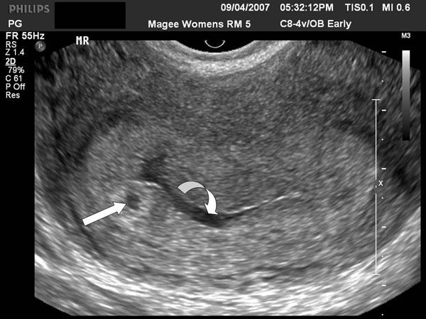
The gestational sac can be seen in:
- 4 weeks and 3 days from the first day of the last menstrual period on TV ultrasound and is usually 2–3 mm in diameter; nine0028
- 5-6 weeks from the first day of the last menstrual period on TA ultrasound, and its diameter is about 5 mm.
Calculation of term by the size of the ovum:
Menstrual gestational age = Mean diameter of the fetal egg (mm) + 30 or 35 (if the diameter is less than 16 mm)
For example, the average diameter is 5 mm, gestational age = 5 + 30 = 35 days, or 5 weeks.
Normal growth rate
Fertilized egg grows by an average of 2 mm every 2 days from 4th to 9th-th week of pregnancy, but such indicators are usually not used to determine the progress of pregnancy, but are used only to confirm the diagnosis of its regression (fading).
What to look for
If no embryo can be detected with a gestational sac of 16–24 mm, suspect a miscarriage or an empty gestational sac and repeat the ultrasound in a week.
The size of the fetal egg is more than 25 mm and the absence of an embryo speaks in favor of a pathological pregnancy (frozen, empty fetal egg). nine0003
If the size of the ovum decreases in the presence of a live embryo after 9 weeks, oligohydramnios can be suspected, although amniotic fluid level is usually not performed until 18–20 weeks.
Yolk sac
Before the embryo is born, the yolk sac, an important structure of the fetal egg, can be seen by ultrasound. The yolk sac is a 100% confirmation of the presence of a uterine pregnancy. With an ectopic pregnancy in the uterine cavity, you can find a formation that is somewhat reminiscent of a fetal egg, which is a specific reaction of the endometrium to pregnancy. nine0003
The yolk sac is located between the amnion and the chorion (two formations of the fetal egg, from which the fetal membranes and the placenta are formed) – in the chorionic space.
Normally, with an average diameter of the ovum of 5 mm, the size of the yolk sac is up to 6 mm (on average, 3–5 mm). The maximum size of the yolk sac is observed at 10 weeks and is 5–6 mm. During the laying and development of the organs of the embryo, the yolk sac is partially used in the formation of the intestine.
Amnion
Amnion is a shell (membrane) inside the ovum that contains the fetus. You can consider the fetal membrane up to 12 weeks of pregnancy with a fetus size of 5–7 mm. The completion of the formation of shells occurs by 12–16 weeks.
Coccyx-parietal size (KTR, CRL)
The coccyx-parietal size is the greatest length of the embryo, but significant errors are possible in measuring the size of the embryo. In the short term, when the limbs are poorly visible, it is more difficult to see the coccyx. By measuring this size of the embryo, a more definite gestational age can be set – up to 4 days. nine0003
With ultrasound, an embryo measuring 1–2 mm can be seen with a gestational sac measuring 5–12 mm at 5–6 weeks of gestation. During the day, the size of the embryo increases by 1 mm. A difference of 5 mm between the size of the embryo and the fetal egg is considered the minimum norm for pregnancy.
Calculation of gestational age
- Menstrual period in weeks = CTE (cm) + 6 (if CTE is less than 1 cm)
- Menstrual period in weeks = CTE (cm) + 6.5 (when CTE is greater than 1 cm)
- Menstrual period in days = CTE (mm) + 42 (with CTE up to 84 mm)
Embryo heartbeat
Embryo/fetal heartbeat can be detected at 6 weeks of gestation with embryo sizes greater than 2 mm, which is a reliable sign of a live pregnancy. However, in embryos up to 5 mm (TV ultrasound) and 9 mm (TA ultrasound), the heartbeat may not be noticed, therefore, in such cases, it is advisable to repeat the ultrasound after 3-5 days.
Before 6 weeks of gestation, the fetal heart rate is 100-115 beats per minute. Within 6–9th weeks, the frequency increases and reaches a maximum at 8 weeks – 144-159 beats per minute. Starting from 9 weeks, the frequency slowly and slightly decreases.
Slow heart rate is called bradycardia . However, it can also be a sign of fading pregnancy.
Bradycardia is set if:
- heart rate is less than 80 beats per minute with CTE less than 5 mm;
- less than 100 beats per minute at CTE 5–9 mm;
- less than 110 beats per minute at CTE 10-15 mm. nine0028
Collar zone (collar space, OT, NT)
Collar zone is an accumulation of lymphatic fluid between the skin and soft tissues of the embryo behind the neck (in the collar region), the thickness of which is a prognostic indicator of chromosomal abnormalities of the fetus. It is measured at 11-14 weeks (up to 13 weeks 6 days) – this is part of the first trimester prenatal genetic screening, which also includes the determination of biochemical markers in the mother’s blood. nine0003
Since the size of the neck crease depends on the size of the embryo, it is very important to measure within the limits of the gestational age – then the measurements will be not only accurate, but also of practical importance. That is, they do it with a CTE of 45–84 mm (11–14 weeks). Such a strict framework for taking measurements is associated with the peculiarities of the anatomical changes in the developing embryo, especially the lymphatic system and the exchange of lymphatic fluid.
Normal dimensions of the VZ are up to 3 mm. Pathological are considered sizes of 5 mm or more at 16-18 weeks, 6 mm or more – at 19-24 weeks.
In contrast to the nuchal space, which is measured in the first trimester, the thickness of the neck roll is measured in the second trimester . The neck roll is the distance between the outer edge of the occipital bone and the skin of the fetal neck. In many cases of chromosomal anomalies, especially those accompanied by malformations of the cardiovascular system, there is a violation of lymph metabolism and it accumulates in the region of the neck roll. Measurements are taken at 16–20 weeks (usually as part of an anatomical ultrasound), and measurements greater than 6 mm are abnormal. nine0003
Nasal ossicle
The presence or absence of the nasal ossicle and its length are recognized as markers for trisomy 21 (Down syndrome). Length is measured at 11–14 weeks as part of prenatal genetic screening. The absence of a bone or its length less than 2.5 mm may be signs of Down syndrome.
There are other ultrasound signs that are used by specialists to clarify the normal development of pregnancy or abnormalities, including miscarriage, empty gestational sac and others. nine0003
Starting from the second trimester (after 14-16 weeks), all measurements of certain parts of the fetus, as well as various indices that are calculated based on these indicators, are plotted with initial percentile curves (95%, 50%, 5% ), which were obtained by statistical processing of data from numerous measurements of fetuses in the past to determine the normal growth and development of this fetus. Creating individual graphs of important ultrasound indicators allows you to see the development of the child in dynamics and evaluate it more correctly. nine0012
It is always important for pregnant women, most of whom do not have a medical education, to know whether the gestational age according to ultrasound coincides with the expected one according to menstruation or the day of conception (not necessarily accurate to the day and hour), where the fetal egg is located, including in relation to the walls of the uterus (bottom, posterior wall, anterior wall, internal pharynx), how many fetal eggs and embryos are there, whether it is a live pregnancy or it has faded.
However, it is very important not to cross-check these results yourself with data from numerous sites, because the lack of the ability to correctly interpret these data will lead to false conclusions and unnecessary stress. nine0003
Remember: a healthy pregnancy does not need multiple ultrasounds and frequent monitoring!
 youtube.com/embed/88Ds6b24T9U” frameborder=”0″ allowfullscreen=””>
🔍 Popular issues about pregnancy and answers to them
Yolk bag
2302 Related
April 25, 2018 I have a second long-awaited b. After today’s ultrasound, she’s not herself. Was on ultrasound to confirm the fact b weeks ago, put 4 weeks. b. Next visit in a week to detect heartbeat (today). Deadline ALREADY (IN A WEEK) 6 weeks. and yolk sac 3 mm. The doctor says: I don’t like that he is so small (yolk sac). I rummaged through the entire Internet, for 5-6 weeks the norm is 3mm. It seems to have calmed down, but doubts remain. Can anyone tell me if this has happened and what are your thoughts? nine0003 0 11298
Yolk sac 1.1 mm!!! Is there any way to save the pregnancy?
Complain
14 June 2015 07:52 at Personal journal HCG on the 11th of 1713 … PJ 7 mm. According to my calculations, 24 days have passed since ovulation. I ALWAYS FEEL IT!!! the obstetric term is set at 4 weeks and 6 days, and in terms of size PYA-5 WEEKS! and 0 days! THOSE. Did you ovulate before your period? I don’t understand… Although she was in this cycle! I’m sure! I will not confuse such pain! I began to feel it after the cancellation of OK! So, according to my calculations, the embryonic period is 3.5 weeks! This is important for me, because yesterday I was very scared! They said the size of the yolk sac is too small … 1.1 mm! and should be 2-3 mm. They even canceled the tests (((. I see, the doctors don’t believe that B will continue to develop. I was told to hope for the best! So I think, VM is visible from the 6th week. And I have 3, …
0 134564
1st ultrasound after eco. No yolk sac
Complain
Hello! I really need your help. Yesterday I was on my 1st ultrasound (24 DPP 5 days). Two fetal eggs, one very small, they said that they most likely would not develop, and the second was larger – 7 mm long and 6 mm wide, but the yolk sac and the embryo is not visible.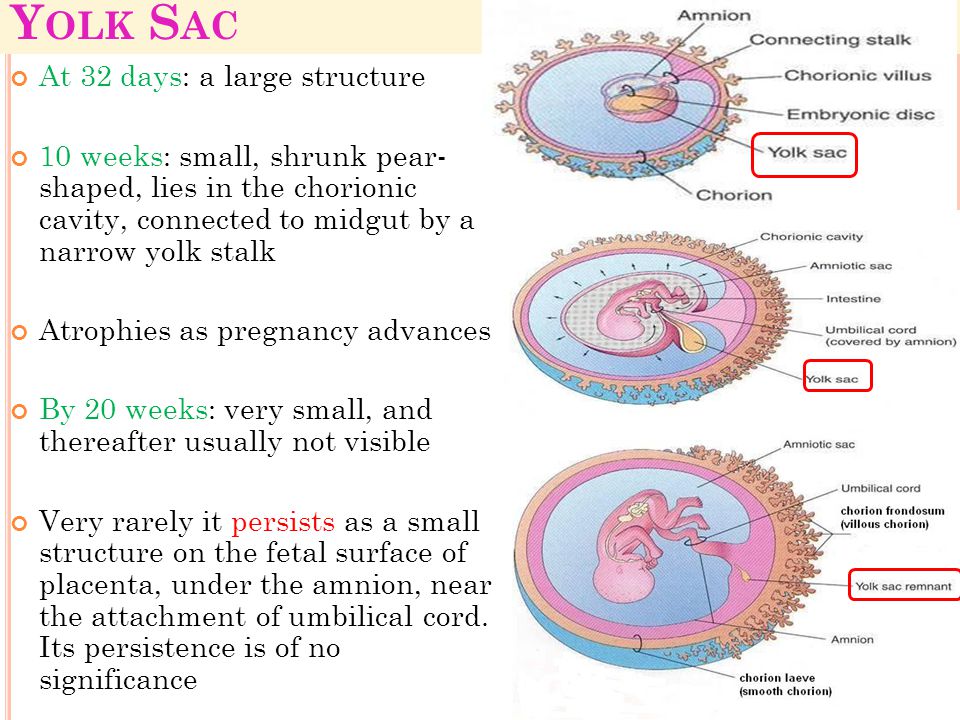 I became hcg – 5500. They said to come in a week. Do you think there is a chance that it will still appear, or is that all? We have been waiting for this miracle for 8 years! If someone had this, and then everything turned out write, I’ll wait
13882
Uzist girl mixed up the yolk sac with the embryo
As prescribed by the doctor, I came to the ultrasound to track the heartbeat (short term, 6.5 weeks). The uzistka turns the screen on me and asks: what do you see? I say: two balls, are there two? Yes, he says, there are two, but don’t flatter yourself, one can dissolve, and so on.
She asked about the heartbeat, about whether both of them can see it. Yes, he says everything is fine.
And then, when I was already dressed, she told me: you know, I got it wrong, it was not an embryo, but a yolk sac. And the heartbeat in it just echoed 😳
I fell out.
I ask the doctor: how is it?
It happens, he says, is duplicated …
And she appointed me another ultrasound in 3 weeks to look at the dynamics of development. Study…
0 10477
Yolk sac
Complain
19 March 2019 04:32 at Is it true?? Could it be? I still can’t figure out where the second yolk came from the next day after the ultrasound ??? Was one big became two small?? And I still don’t understand why they put 6 weeks and 5 days on March 16 and put 6 weeks and 1 day the next day🤷♀️🤷♀️🤷♀️
0 15172
Anembryonic, short term or mole?
Complain
28 August 2020 19:26 at Personal Journal
Good evening.
PM 12.07. ovulation was somewhere around 25-27.07.
Hcg 13.08 – 497
Ultrasound on August 25 shows an empty fetal egg 16 mm, no yolk sac, no embryo. The term according to ultrasound is 3.5-4 weeks. Anembryony.
Hcg 27.08 – more than 16,000.
I read on the forums that they find an embryo, you just have to wait. Confused by the lack of a yolk sac. I don’t know what to think anymore. At first there was hope, now it is less and less. I did an ultrasound today – the same. I’ll wait another week and redo the ultrasound again. Would like to hear opinions. The doctor says you can wait. He says it doesn’t look like a bubble drift. nine0003 0 11378
Uzist woman confused the embryo with the yolk sac 😳
Complain
17 March 2019 12:12 at Questions during pregnancy
As prescribed by the doctor, I came to the ultrasound to track the heartbeat (short term, 6.5 weeks). The uzistka turns the screen on me and asks: what do you see? I say: two balls, are there two? Yes, he says, there are two, but don’t flatter yourself, one can dissolve, and so on.
She asked about the heartbeat, about whether both of them can see it. Yes, he says everything is fine.
And then, when I was already dressed, she told me: you know, I got it wrong, it was not an embryo, but a yolk sac. And the heartbeat in it just echoed 😳
I fell out.
I ask the doctor: how is it?
It happens, he says, is duplicated . ..
..
And she appointed me another ultrasound in 3 weeks to look at the dynamics of development. Not registered st…
0 13337
problem of small towns
Complain
May 12, 2014 03:47 pm at Personal Journal
I’ll start with the second baby. And the third pregnancy. The last one turned out to be anembryonic. Here we are 9 weeks old, and there is still no confidence in my head that everything is going as it should. I cannot even consider myself pregnant until I am convinced that there is life in me. Just after the previous time, the feeling of fear that everything is repeated stubbornly stuck in the mind. Another thing is that the town is small. And there is nowhere to do an ultrasound here (so they told me in our clinic), although my seller says that she did it last week. I so understand our gynecologist not strongly wants to bother. At six weeks I went to the neighboring area for a paid ultrasound for myself. She left with nothing. Just hearing about my previous frozen, the uzistka is no more than m …
0 20514
Small embryo size
Complain
March 9, 2015 9:29 pm at We are 7 weeks obstetric. I had an ultrasound today, they wanted to hear a heartbeat. But the result is not very … The size of the fetal egg is 24 mm, the size of the yolk sac is 5 mm, but the CTE of the embryo is only 3.7 mm. We didn’t really hear the heartbeat, although my husband and I could see on the screen that the embryo was pulsing. (Or really wanted to see it). The doctor said that the size of the embryo is small, does not correspond to the obstetric period, and so far she cannot say anything for sure, she needs to take hCG and again in 10 days for an ultrasound scan. I’m upset, I’m sitting worried. I try to believe in the best. Husband calms down. Has anyone had anything similar? What ended? And our photo
0 281133
Small CTE. Let’s have positive stories)
Complain
May 5, 2016 18:53 at I want a child
Girls, hello everyone!
We went for an ultrasound today, we can’t hear our heart (the last menstruation was on 03/16/16. Embryo transfer on 4/4/16. The diameter of the yolk sac is 3.6 mm, the structure of the chorion is 3.2 mm, the heartbeat is not detected, hypoplasia of the chorion.
I want to hear positive stories from you.
And then I was upset about something, the doctor said a very small embryo, it is lagging behind, there should already be a heartbeat. nine0003 1 141194
Small CTE and no heartbeat
Complain
March 7, 2013 2:24 pm at Issues during pregnancy 4 days. The conclusion of the ultrasound examination is that the fetal egg is deformed (it is oval in shape: length 1.8 cm, width 0.5 cm), hypertonicity, the yolk sac is present, the corpus luteum is also present, the CTE of the fetus is only 2.7 mm, there is no heartbeat. They wrote that this corresponds to 3 weeks, but I already have 5, 5
2 weeks ago I had an ultrasound and they saw only a fetal egg without an embryo. HCG passed today – I’m waiting for the result
They said to wait. .. It’s too early to get upset, it happens…
Has anyone had the same fetal growth retardation? Is there still hope? nine0003 0 166231
Who had a small PU with an Embryo and a heartbeat?
Complain
9 December 2014 12:47 at Questions during pregnancy not very informative). I put it on an ultrasound.mm is written), I see the embryo (!!! Hurray !!!!!), CTE 2.6, there is a good heartbeat. In general, the doctor is all happy and I, along with her (we have a long joint struggle with the reluctance of my body to get pregnant) In general, he sets the term for KTR 6 + 1. I did all my work, I come home, I take out a conclusion with photos and to …
0 601669
A small fetal egg for an embryo!
Complain
October 24, 2016 12:35 pm at ktr 5.3mm; the yolk sac is 4.6 mm, the chorion is 2 mm (4 days ago on another ultrasound, the chorion was 3.5 mm), the embryo corresponds to a period of 6 weeks and 2 days. Heart rate 106 beats per minute. Conclusion – chorionic hypoplasia, oligoamnion, risk of non-developing pregnancy. The uzistka said that 100% will freeze, she doesn’t advise saving everything, she won’t survive early, there will be chromosomal abnormalities. nine0003 0 149
Our little MIRACLE, which we still can’t believe in
Complain
27 March 2018 15:01 at Personal journal
Hello girls! I want to tell you about our Miracle baby. We went to her for a very long time, for 10 years, we went through a lot, there were 4 IVFs (during the last IVF, it was in November, we planted 2 five-day-olds, I saw striped tests from 4 dpo, but in the end my ddimer went off scale and babies mine couldn’t develop further, clexane didn’t help), then I made an appointment with Wagner (hematologist), my appointment was supposed to take place in 1.5 months, plus I decided that from the new year I would start collecting documents for IVF again and somehow I turned off my thoughts, although I want to say that I never really strained, I believed that everything would work out with my husband. One fine night, I feel very bad, dizzy, me …
18 61696
Passed hormones)))
Complain
28 August 2015 20:50 at Personal journal )) I went to take it for a fee, I’m waiting for the results, I’m worried. 10 days ago there was very low hCG, yesterday there was a very small yolk sac on the ultrasound, which does not correspond to my term, but the embryo is visible, it floats, they prescribed utrozhestan for the night and magneB6. I hope all will be ok!!! nine0003 0 7223
May 16, 2021 11:23 am at Personal Journal
Complain
Good day everyone!
I’m worried about the small yolk sac 🙁
She was on ultrasound at 8.3, pya 35mm, ktr 18mm, heart rate 174 beats / min, press 2.2 mm, jt 17mm.
I paid attention to the press when I already came home (
In the past, the ultrasound was at 6.3, the press was 2.7. But then ultrasound was performed transvaginally, and this time transabdominally. Could this be the reason for the error?
I would be grateful for any information. On the Internet, it is somehow scarce (and mostly scary).0 7224
Some kind of mystic!!! First twins, then triplets…
Complain
December 30, 2018 11:00 am at Personal Journal
Hello girls. I will share my story. This is my second pregnancy. The first, unfortunately, ceased to develop for a period of 7-8 weeks. Then for the first time I went for an ultrasound at 6 weeks. In one fetal egg, 2 yolk sacs were found (twins would be expected). They advised me to come back in a week. I came a week later and two embryos had already appeared on the ultrasound. But they were very small and did not correspond to the deadline. One was found to have a pulsation, and the other did not. We were advised to wait another week. A week later, she came and both babies stopped developing … Hysteria, pain, tears, purge … Then they made a vacuum aspiartion. After this incident, I passed a lot of tests: for hormones, infections . ..
..
4 26468
Ultrasound 8-9 weeks pregnant
Complain
July 6, 2019 13:25 at Personal journal At 8 weeks and 3 days (obstetrics) she did an ultrasound, the uzist said everything was fine, the gynecologist was silent, but she hadn’t registered yet, she scheduled an appointment for July 9. I’m sitting reading Uzi:
Inner dia.fruit.egg 28 mm
Ktr 16 mm
Yolk sac 2.8 mm.
Is it not a small yolk at such a time, and a fetal egg? I read on the Internet that the yolk should be at least 4.0, and the fetal egg should be at least 2 times larger than the ktr of the fetus .. Yes, and I compare it with Uzi with my son at the same time:
Inner.dia.fruit.egg 34.8 mm
Ktr 15.7 mm
Yolk sac 4.9.
I’m very worried, before my son there was a ZB at this time, and a small yolk one shows how much the child has enough food .. What are your sizes for ultrasound with such a period?
0 12559
The story of our MIRACLE!!!
Complain
29 December 2017 14:28 at I want a baby Close girlfriends from the baby know my gloomy history of previous pregnancies. I’ll tell you briefly. The first pregnancy unplanned occurred with PPA in the spring of 2016. Then I didn’t really know what ovulation was, I never tracked it, I didn’t even follow the cycle) There was a delay of several days, I started to smear, I took it for menstruation, but they passed 2 days, then no and no. At 9-10 day delay, I decided to take a test, there was a pale line, I thought the result was negative, I was sure that it was positive when it was like with ovulation tests, a greasy fat line))) Then another test the next day, the line is already obvious. I surrendered hCG, Ros …
249 1723581
My sad 😔 experience …
complain
August 11, 2017 20:48 Personal journal
I want to save information for myself .. I clearly remember. Let me start by saying that I am only 20 years old. I got married at 18 (with my husband, you can say from childhood .. from the age of 13), it was necessary to give birth urgently, as the doctors were afraid, they said either it was necessary to give birth early or not to give birth. I have a history of many diseases, there are those that say that I am forbidden to get pregnant. (Hemorrhagic vasculitis of the skin-articular-abdominal form, this is an autoimmune disease), which later can cause antiphospholipid syndrome and all sorts of bad things, but that’s not the point … my husband also has an autoimmune disease (autoimmune thyroiditis, hypothyroidism, Schamberg’s vasculitis), it’s essentially dangerous for us to have children but I really want my own. We started planning…
3 342048
Retrochorial hematoma …
Complain
July 1, 2020 13:16 at ), well, a week later, I started looking for a good gynecologist, since there was a huge queue for mine at that time. Found. PYURBEEVA Elena Nikolaevna Clinic “Intramed” on Savushkina (St. Petersburg), the clinic specializes in gynecology and urology. The doctor (as it is written) is a candidate of medical sciences, a doctor of the highest category (((came, a friendly person. She examined, began to do an ultrasound. On the monitor there is a round ball (fetal egg), inside which is a vesicle (yolk sac), and next to a curve of an incomprehensible shape is a substance …..(((As I understood right away (far away in medicine) that this is a fetal egg and a yellow sac, and next (du …
1 7378
Long-awaited, but miss
Complain
April 27, 2016 11:02 am at Personal Journal
Girls… I’m 28. I’m married. We have an apartment that we have already managed to buy out in a mortgage and a car for which we have almost paid off. But there is no happiness. There is no small and native happiness. My husband and I have been trying to get pregnant for almost 2 years. They went from doctor to doctor. As a result, I was given a pituitary adenoma in question. The point was that according to all the ultrasounds I had perfect ovulation … but as it turned out, prolactin was almost 2000 … But when taking Dostinex, it fell below 10 in a month and a miracle happened. I saw the long-awaited 2 stripes … Happiness was limitless, I did tests every day and we were touched, seeing that the stripes were getting brighter and brighter. I gave hCG in 2 days: 33,216,413,826 … on an ultrasound on Friday they saw a fetal egg in ma …
1 15415
Today
Complain
January 6, 2015 9:28 pm at Personal Journal
Honestly I didn’t even know what to call the post. And so my nerves were on edge … I decided not to wait for Jan 8th. The state of uncertainty is killing me (I went to donate blood for hCG and signed up for an ultrasound in Demeter. And so is my ultrasound. I decided not to tell the uzistka about my situation … if I had told immediately about my diagnosis, she would immediately look for a missed pregnancy. It all started well, the egg is in the uterus, the fetus is 6-7 weeks old, something else was good (but I was interested in the heartbeat) and now we got to it, but she didn’t see it (((By the way, they confirmed the tone of the uterus! I also couldn’t find the yolk sac. I didn’t cry anymore, after all, I’ve already heard this for the third time ((She asked me: Do I have other ultrasounds? And I suggest going to …
1 991629
the embryo was not visualized…
Complain
August 18, 2014 11:30 am at Personal journal Foliber, vitamin E, and utrozhestan were prescribed. Now we have a second attempt, the first pregnancy was in 2013, just before the New Year, for a period of 3-4 weeks, we lost the baby, cleaned it and sent it home. I’m afraid for the baby, in principle, nothing really bothers me, there are no strong pains, it doesn’t bleed, they pose a threat, because there were problems from the first, according to the analyzes, all the results will be told in the next week I just have a second visit to the doctor. Nausea does not go away, almost all the time, sometimes it just leaves you alone, you are madly hungry to sleep, but you still have to work in the usual rhythm, in principle, the work is not difficult, ri . ..
..
0 402008
Collection closed! Let’s help Sasha Bednyagina survive!!!
complain
June 25, 2015 14:13 In we need help for children
http://www.baby.ru/post/407066577-321011550/ Bednyagina Sashenka, 24.10.2012 Diagnosis of Herminogenic malignant malignant malignant maleby yolk sac tumor Collection to the German clinic “Uniklinik Düsseldorf” (uniklinik-duesseldorf.de/hyperthermie), a new invoice received from the German clinic in the amount of 70,000 €, this is for two courses of hyperthermia and a preliminary calculation for the operation. The topic is opened by volunteers http://www.baby.ru/u/usr2003424/ and http://www.baby.ru/u/usr
7/
Sashenka – our sun was born on time, 10/24/2012, weight 4090 kg, height 54 cm, grew and developed even ahead of her peers. Fighting character showed from birth. Everything was fine, the doctors wouldn’t have any complaints either …
286194
My first
Complain
June 28, 2017 11:32 am at everything is not going the way we want . .. I met my future husband at the age of 17, we met him for 16, 5 years, in 2010 we officially became husband and wife and began to live together. Like many girls, I had my own life plan – meet, get married, go on vacation, live for yourself… but we assume, but God disposes. I am simultaneously treated for rest, I drink antibiotics, as a result, the thrush recedes, but not for long 🙁 due to my stupidity, I missed the moment of simultaneous treatment of my husband with m …
3 16342
Ultrasound at 5 weeks and 3 days (obstetric period).
Complain
3 June 2010 12:09 at Women’s consultation
I did an ultrasound in vitro. egg 11mm, embryo 2mm, yolk sac 1.4mm. Cystic corpus luteum 23 by 18. It turns out that the fetus is an egg for 6 weeks. more than necessary????? The embryo corresponds to the deadline, thank God. Pulsation +. But the yolk sac is small (((. So the bead lacks something and it suffers? Or how? It was then 22-23 days after ovulation.
0 1717
5 weeks and 6 days)) ultrasound
Complain
February 1, 2015 19:22 at Personal journal
Went for an ultrasound today! I liked everything very much, they measured everything up and down, even measured the blood flow in modest arteries))) I didn’t know that they were doing it in such a short time.
But the most important thing is that I heard and saw my baby’s heart))) heart rate 99 beats per minute, at first I thought that it was not enough … I changed the entire Internet, the norms are 4-6 weeks 80-85, 6 weeks 100-130 … so we just fit into the norm)). Given that 4 days ago neither the embryo nor the yolk sac was visible, the heart had just begun to beat.
Well, the main dimensions are svd 14.8 mm (4 weeks from conception), Villous Horton annular, thickness 4.3 mm,
Yolk sac 4.5 mm,
Ktr 2.8 mm,
Heartbeat +, heart rate 9nine,
En…
0 4441
Made an ultrasound
Complain
August 18, 2015 11:30 am at Personal journal
Immediately warned that ovulation was late.
In total, they wrote that there is a fetal egg 6 mm, the embryo is not visible, there is no yolk sac either. Yellow body 19 mm. There is no tone, no detachment. They put a uterine pregnancy, 4-5 weeks. Girls, why is there no yolk sac? As I understand it, it should be ….. Is the corpus luteum not small? And I still don’t understand, if there is no detachment, then what was smeared at 18 DPO ???? nine0003 0 23415
Panic after ultrasound 9 weeks 3 days
Complain
October 23, 2015 19:01 at Personal journal
Girls, I’m sitting sobbing!!! The clerk said everything is fine, don’t worry. (I’m standing after the ST) Here are the readings of the PJ 28 * 11 * 25 Yolk sac 2.7 mm Embryo ktr 22 mm Chorion annular 6 mm, homogeneous The pulsation of the heart tube is distinct But I looked in the internet – the thickness of the chorion should be 7 – 9 mm Yolk sac also very small …. Please write your testimony for this period
1 22776
This page contains the most popular posts and comments of our users on the topic “Little yolk sac”.