Cyst vs tumor in dogs: Dog Tumors: Signs, Types, Diagnosis & More
Dog Tumors: Signs, Types, Diagnosis & More
To keep the lights on, we receive affiliate commissions via some of our links. Our review process.
Fear sets in when you feel a lump or bump on your dog that you haven’t felt before. Thoughts of your dog having a tumor take over. But keep in mind that growths and masses are common with dogs, and not all dog tumors are cancerous.
>We’ll help answer some of your questions about the signs, symptoms, and types of tumors in dogs, as well as what to expect when you see your veterinarian — a prompt vet appointment is important with any abnormal mass or growth you notice on your dog.
Table Of Contents
- What Does A Tumor Look Like On A Dog?
- What Are The Symptoms That It Might Be Cancer?
- Types Of Dog Tumors
- How Do Vets Diagnose Dog Tumors?
- Cutting The Cost Of Cancer Treatment
- Learn More About Dog Lumps And Bumps
What Does A Tumor Look Like On A Dog?
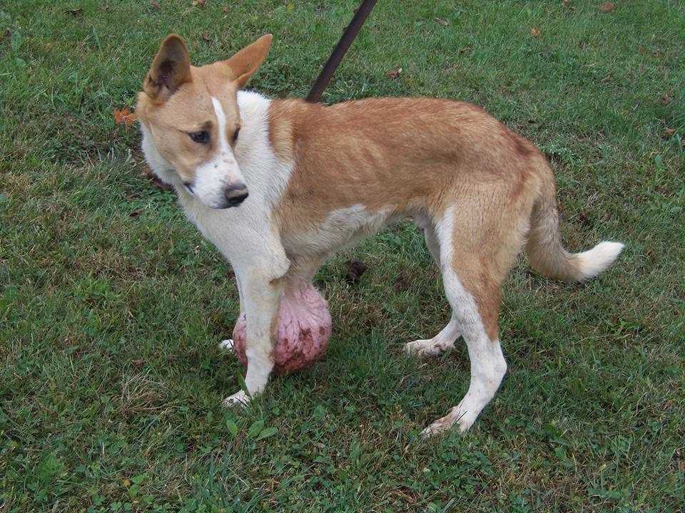
Tumors come in all shapes and sizes and can be benign or malignant (cancerous). These are the most common signs of tumors that you can detect on your dog. (Some dog tumors are internal, so you won’t be able to see or feel them.)
- A raised area on the skin or under the skin
- Can be round or have irregular borders
- Can be red or darkly-pigmented and also may bleed
- Size can range from pea-size to a much larger mass
- Can be soft or hard to the touch
Dog Cyst vs Tumor: How Do I Tell The Difference?
Cysts are fluid-filled sacs under the skin that are usually easy to move around, while tumors are typically more solid. A cyst also may drain a white, yellow, or green discharge. But it can be challenging to determine the difference. The only way to know for sure is by seeing your veterinarian.
What Are The Symptoms That It Might Be Cancer?
Several signs indicate that your dog may be dealing with cancer. It helps to be aware of cancer’s physical and behavioral signs, especially if the tumor is internal.
- Large growths
- Growths that are constantly growing and changing
- Abnormal swelling in the body
- Enlarged lymph nodes
- Sores that won’t heal
- Loss of appetite
- Weight loss
- Lethargy
- Strong odors
- Coughing or difficulty breathing
Learn more about these cancer signs in this video:
Types Of Dog Tumors
There are many different types of tumors in dogs that can occur in any area of the body, but here are some of the most common.
Mast Cell Tumor
Mast cell tumors are one of the most common malignant tumors in dogs. They most commonly grow on the skin but can also form in the internal organs. The most common locations are the limbs, chest, and lower abdomen. This type of cancer produces tumors that release the chemical histamine, which causes the tumor to be red and itchy.
These tumors originate in the mast cells and can range from low grade, where treatment is to surgically remove the tumor, to high grade, where the tumors have spread, and treatment goes beyond surgery to also include radiation and chemotherapy.
Lipoma
Lipomas are a very common benign fatty tumor that feels soft to the touch and can be moved around under the skin. This type of tumor usually isn’t a problem and is only removed if it’s bothersome to the dog. In rare cases, lipomas can be a malignant tumor called a liposarcoma.
Histiocytoma
Histiocytomas are benign tumors that mostly affect dogs under three years old. These “button tumors” are hairless, red, raised lumps that often resolve on their own within a few weeks. But some can grow quite large rapidly and become irritated, so your vet may recommend removal if this occurs.
Melanoma
Melanoma is as much of a risk to dogs as it is to humans, although there are both benign and malignant forms. These tumors in dogs occur most often on the lips, mouth, skin, eyes, or nail bed. This tumor appears as a dark spot but can sometimes be pink, and it can be either raised or flat. Malignant melanomas can spread quickly, requiring a combination of surgery, radiation, and immunotherapy.
Lymphoma
Lymphoma is a cancer of the lymph system and is often first noticed in swollen lymph nodes behind the knees, under the shoulders, and in the jaw. A swollen lymph node can feel like a fairly hard lump that moves easily under the skin. Chemotherapy is the most effective treatment for lymphoma in dogs.
Osteosarcoma
Osteosarcoma is a painful malignant bone cancer that most often affects dogs’ legs but can occur in the hips, pelvis, or jaw. Signs include limping, swelling, and general lethargy. It’s more common in larger breeds than in small dogs.
Treatment typically includes amputation followed by chemotherapy. Sometimes amputation can be avoided by bone grafting or metal rod placement once the dog tumor is removed. This can be costly and invasive but may be worth it when considering how your dog will deal with amputation.
Mammary Gland Carcinoma
Mammary gland carcinomas are tumors that grow in the mammary (breast) tissue. They occur most frequently in female dogs, especially those who aren’t spayed or weren’t spayed until after their second heat cycle. They can feel like firm, nodular lumps underneath the skin on the abdomen, often close to the nipples. Not all mammary gland carcinomas are malignant, but surgery is required to determine whether it’s cancerous or not.
Hemangiosarcoma
Hemangiosarcomas are most often cancerous tumors on a dog’s spleen, heart, or skin. This cancer grows in the cells that line blood vessels, so there’s a high risk of rupture, which causes internal bleeding. It’s often treated by removing the tumor and then prescribing a chemotherapy regimen to reduce the risk of the cancer spreading.
Papilloma
Papillomas are benign tumors caused by a virus known as papillomavirus. They look like warts and appear around the eyes, on the lips, and inside the mouth. It can take weeks or months for these warts to go away, and your dog may be contagious during this time. Treatment is often just trying to make your dog more comfortable but may also include removal.
How Do Vets Diagnose Dog Tumors?
Your vet will perform a physical examination to determine whether your dog’s growth is a cyst, some other type of skin growth, or a tumor. In the case of internal tumors, blood work and other diagnostic tests can often point toward an organ that needs closer examination.
However, vets can’t definitively diagnose a dog tumor without taking a sample to inspect under a microscope. Sampling involves either a fine needle aspiration or a biopsy, and in some cases, surgery is required to obtain a sample.
Cutting The Cost Of Cancer Treatment
If you’re worried about the cost of treatment, you may be thinking about how to treat dog tumors at home. Though there may be some holistic ways to try and treat noncancerous tumors on dogs, that’s not the best choice. You need to see your veterinarian to determine what kind of tumor your dog has so they can recommend the best treatment. The best way to defray the high cost of diagnostics and treatment is by having pet insurance.
Pet Insurance Claim & Story
Below is a real-life pet insurance claim from a Healthy Paws customer.
- Pet: Dixie Mae (7-year-old American Staffordshire)
- Diagnosis & Treatment: 23 growths removed, not all benign
- Total Vet Cost: $15,880
- Plan Options: 80% reimbursement with $500 deductible (some pre-existing conditions not covered)
- Reimbursement: $9,577
“A couple of months ago, I noticed a small bump on her backside to the right of her tailbone,” says pet parent Susan. When the growth began to get bigger, they decided to get it checked out. The vet decided to remove the growth, but before that happened, a few more growths became noticeable. “It ended up that three of the four growths were malignant and the other one was a benign growth.”
Sometimes, when you find a growth on your dog, it can be a simple procedure to remove it. However, that’s not always the case. You may find that you need to have tests, biopsies, and surgeries to deal with the situation.
Susan says, “Given Dixie Mae’s history of skin issues, I have come to realize it is better to be proactive than reactive.” Susan inspects Dixie May regularly to stay on top of growths that arise. Over the past two years, she has had 23 growths removed, some malignant and some benign. “Having Healthy Paws Pet Insurance has enabled me to truly be proactive and have Dixie treated by the best veterinarians,” says Susan.
Learn More About Dog Lumps And Bumps
We all want to make sure we keep our pups happy and healthy.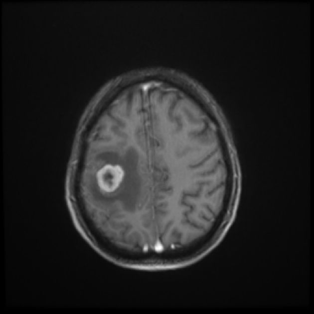 So remember, if you see or feel something abnormal, it’s always best to get your veterinarian to check it out. Dogs can develop so many different types of lumps on or underneath their skin. Some are harmless, but others require prompt treatment. Being informed and prepared will help you get the best care for your precious pup.
Tagged With: Aging, Cancer
Tumors, Growths, and Cysts in Dogs
It’s common to find lumps and bumps on all types of dogs. Growths, tumors, cysts, and masses can appear on dogs at any age, but they are among the most common health issues seen in older dogs. As a dog owner, it’s helpful to understand the different types of growths you may encounter. Any persistent, unusual mass, or growth should prompt an immediate call to your veterinarian.
What Are Tumors, Growths, and Cysts?
Most veterinarians will call any unknown lump or bump a growth, mass, or tumor. In general, the terms can be used interchangeably, but most vets avoid the word tumor unless the mass has been determined to be a type of cancer.
The Spruce / Kelly Miller
Symptoms of Tumors, Growths, and Cysts in Dogs
Abnormal growths can occur anywhere on the body or in the mouth. Warning signs include:
Symptoms
- An abnormal skin lump or a bump ranging in size from very small to very large
- A swollen area (particularly within the body)
- An oral growth
- Enlarged lymph nodes
- Lameness or swelling affecting a bone
Abnormal Lump or Bump
- Sebaceous cysts, adenomas, and adenocarcinomas are common types of skin cysts that contain sebum, a thick, oily material normally found in the skin around the hair follicles. These masses may be found anywhere on the body. Sebaceous cysts are benign but can also be mistaken for a malignant tumor called a sebaceous gland adenocarcinoma or a benign mass called a sebaceous gland adenoma.
 If the cyst does not bother your dog, your vet might leave it alone, but a cyst can be surgically removed if necessary. Once removed, the cyst should be sent to a lab so a veterinary pathologist can determine that it is, indeed, just a sebaceous cyst or an adenoma or adenocarcinoma that may require more treatment.
 - Histiocytomas are red bumps that can appear quickly on your dog’s skin and tend to go away on their own over the course of a few months. Although they are benign tumors, some can grow rapidly and really bother your dog. Your vet may recommend the removal of large or irritated histiocytomas. Unlike other common skin masses, histiocytomas are most frequently diagnosed in younger dogs.
- Skin tags on dogs are similar to those humans get. Some can get quite large and pendulous, hanging off the skin by a narrow stalk. Skin tags are benign and are usually not removed unless they bother the dog or get very large and irritated.
- Malignant melanoma can occur on the skin and/or in the mouth and is thought to be caused by sun exposure. Many of these tumors have a black color but not all will look the same.
- Squamous cell carcinoma is a type of tumor that may be caused by sun exposure. This type of cancer can occur on the skin and/or in the mouth. These tumors can have a pink or reddish color and misshapen, “raw” appearance.
- Mast cell tumors may occur as skin bumps or internal tumors. These masses may release histamine when disturbed, which can have a negative effect on your dog’s body. If your vet suspects a mast cell tumor, your dog may be treated first with diphenhydramine to minimize the histamine release. Once the mass is removed, a pathologist will grade the tumor as I, II, or II. This grading indicates how malignant the tumor is and how likely it is to metastasize (spread to other parts of your dog’s body).
 If the cyst does not bother your dog, your vet might leave it alone, but a cyst can be surgically removed if necessary. Once removed, the cyst should be sent to a lab so a veterinary pathologist can determine that it is, indeed, just a sebaceous cyst or an adenoma or adenocarcinoma that may require more treatment.
 Many of these tumors have a black color but not all will look the same.
Swollen Area in the Body
- Internal masses develop within the chest or abdomen, especially in some senior dogs. Internal masses may be found due to the symptoms they create (difficulty breathing or vomiting, for example) or during a routine physical examination. Internal masses may be benign or malignant and are usually definitively diagnosed through a combination of radiographs, ultrasound, lab work, and biopsy. Treatment depends on the location and type of tumor.
- Mammary tumors are more common in female dogs, particularly in those that are not spayed but can sometimes occur in spayed females as well. Though some mammary masses may be benign, many are cancerous. Prognosis improves when the masses are diagnosed and surgically removed when they are small.
- Lipomas are common types of tumors seen in dogs. A lipoma is a benign fatty mass that can be found anywhere on a dog’s body, typically under the skin. They usually feel soft and moveable and rarely cause pain or discomfort for the dog. Lipomas can be surgically removed if they interfere with your dog’s mobility or comfort, grow rapidly, or rupture (causing skin damage). In rare cases, an apparent lipoma is actually a malignant tumor called liposarcoma. Diagnostic testing can differentiate the two.
 Internal masses may be found due to the symptoms they create (difficulty breathing or vomiting, for example) or during a routine physical examination. Internal masses may be benign or malignant and are usually definitively diagnosed through a combination of radiographs, ultrasound, lab work, and biopsy. Treatment depends on the location and type of tumor.
 In rare cases, an apparent lipoma is actually a malignant tumor called liposarcoma. Diagnostic testing can differentiate the two.
What Is a Lipoma?
A lipoma is a fatty tumor just below the skin. It is a benign (non-cancerous) lump made up from fatty tissue.
An Oral Growth
There are many kinds of growths that can develop in your dog’s mouth. Some growths cannot be easily seen but will cause signs like bad breath, trouble chewing, difficulty holding things in the mouth, oral pain, and pawing at the face or mouth. Of course, these signs could also indicate dental disease and should not be ignored.
- Papillomas are warts caused by the papillomavirus. They can appear on the dog’s lips, face, and inside the mouth. Papillomas are benign but very contagious. They can be removed if they cause problems for your dog, but in many cases, they will resolve on their own.
- An epulis is an oral growth that usually forms on the gum tissue around a tooth. Many epulides are benign, but some can be malignant, so further diagnostics are necessary.
- Gingival hyperplasia is a benign overgrowth of gum tissue that may look a little bit like a tumor in some dogs. This excess gum tissue can be removed if it’s affecting the teeth or is bothersome to the dog. The removed tissue may be sent to a veterinary pathologist just to make sure there are no cancer cells present.
- Oral melanoma can occur in the mouth and may be black in color.
- Squamous cell carcinoma and fibrosarcoma are other common types of cancer that can develop within the mouth of dogs.
 Many epulides are benign, but some can be malignant, so further diagnostics are necessary.
Some oral tumors can affect the teeth and bone in the mouth and face. If your dog has an oral mass, your vet will likely recommend putting your dog under anesthesia so a thorough examination and radiographs can be done.
Enlarged Lymph Nodes
Lymphoma is not actually a tumor; it is a cancer of certain cells within the immune system. However, the first sign of canine lymphoma is often an enlargement of the lymph nodes, which can look and feel like tumors.
Pet owners most often notice lumps in the neck area, but they may also be found in the axillary area (armpits), the inguinal area (lower abdomen near thighs), and the back of the knees. Lymphoma is often diagnosed with a fine needle aspiration or biopsy. Chemotherapy is the most common treatment for lymphoma.
What to Do if Your Dog Has Swollen Lymph Nodes
Lameness or Swelling Affecting a Bone
If you notice that your dog is walking with a gait, favoring a leg, or is behaving otherwise lame, it could be a swollen growth affecting a bone that you can’t feel. Regardless of whether it’s a tumor, growth, or cyst, the area is likely tender and your dog is in pain, which requires a visit to the vet for diagnosis.
Causes of Tumors, Growths, and Cysts
As with humans, it’s tough to pinpoint the direct cause of a tumor, growth, or cyst in an animal. However, the environment or an illness is thought to possibly cause skin problems in dogs. Genetics can also play a major role in the development of other types of tumors, growths, and cysts.
Diagnosing Tumors, Growth, and Cysts in Dogs
When a lump has been discovered, your veterinarian will perform a physical examination. If the lump is very new and potentially temporary (like the result of a bug bite or an injection), the veterinarian may recommend a period of observation, but in most cases, they will perform additional diagnostics to determine the type of cells that comprise the mass. This usually means collecting a sample of the material from the mass and analyzing it under a microscope.
A veterinarian typically collects these samples via fine needle aspirate or biopsy. Evaluation of the samples (often performed by a pathologist) can indicate whether the mass is cancerous, and if so, what type of cancer is present.
If your veterinarian diagnoses your dog with cancer, additional diagnostics will most likely be recommended, including:
- Lab tests such as blood chemistry, complete blood count, and urinalysis
- Radiographs (X-rays) that can reveal signs of metastasis or other problems
- Ultrasound, which can offer a better view of internal organs and look for metastasis
- CT scan or MRI, which will help vets get a closer look at the structure of your dog’s tumor and some internal organs.
Some advanced diagnostics and treatments must be performed by a veterinary specialist.
Treatment
If a fine needle aspirate is not effective (or if your vet thinks it’s not the best option) the next recommendation is usually a biopsy. A biopsy is often performed with the dog under general anesthesia or sedation, but local anesthesia may be used instead depending on the size and location of the mass.
The biopsy may be performed by using a special large needle. Or, the vet may cut into the mass surgically. In some cases, the entire mass is removed surgically and sent to a laboratory for identification.
Prognosis for Dogs with Tumors, Growths, and Cysts
If you and your veterinarian can be proactive with the treatment of your dog’s tumor, growth, or cyst, the prognosis is often good. The smaller the tumor, growth, or cyst, the easier it will be to aspirate or remove, and often results in no additional treatments.
How to Prevent Tumors, Growths, and Cysts
Many lumps, bumps, and growths cannot be prevented, but some can. For example, spaying your dog before her first heat cycle virtually eliminates the chances that she will develop mammary tumors.
In all cases, maintain a healthy diet and an active lifestyle for your dog and see your veterinarian at least annually for preventative care. Adhere to a regular grooming schedule and take note of any lumps or bumps that are new. A photo and a written record can help track growth and if you see rapid change, speak to a vet right away.
If you suspect your pet is sick, call your vet immediately. For health-related questions, always consult your veterinarian, as they have examined your pet, know the pet’s health history, and can make the best recommendations for your pet.
Lumps, Bumps, and Cysts on Dogs
The overall health of a dog is often reflected in their skin. Dogs can get lumps, bumps, and cysts from normal aging, or they can be signs of a problem.
There are two major types of lumps and bumps on dogs: malignant (cancerous) and benign (not cancerous). However, you can’t tell the type or severity of a growth just by looking at it. A veterinarian can take a sample of cells to give you a diagnosis and appropriate treatment plan.
Types of Lumps and Bumps on Dogs
Here are several common skin growths found in dogs, along with info on what they look like and what to watch for:
Benign Tumors
Tumors that are benign are not invasive or likely to spread to other body areas.
Histiocytoma
A histiocytoma is a benign skin growth that usually occurs in dogs less than 2 years of age. They are found on the front half of a dog’s body, usually on the head or legs. Rarely, they can be seen in older dogs or on other areas of the body.
Histiocytomas are pink and fleshy but may get bigger and seem more irritated before improving. These tumors usually regress spontaneously over time without treatment and arise from the skin’s immune cells. They can be diagnosed through microscopic examination of a sample of cells from the growth.
Lipoma
A lipoma may show up anywhere on a dog’s body but is common on the trunk and legs. Lipomas come from fat cells under the skin or are found in muscle tissue. They usually develop in older, overweight dogs. They may become quite large or appear in multiple locations.
A vet can diagnose a lipoma by taking a small sample of cells from the growth to look for fat droplets. No treatment is needed, but these should be monitored for rapid changes. They will gradually enlarge with time, and may bother your dog if they’re located in an area that interferes with motion. When lipomas start to bother your pet, you can consider surgical removal.
Papilloma
A papilloma in young dogs is a contagious, wart-like growth that usually occurs in and around the mouth. In older dogs, they might be seen around the eyes or on other areas of the body. Papillomas are caused by a virus that can be spread through direct contact with an infected dog or contaminated items, such as toys or feeding bowls.
They appear small, fleshy, and round with a cauliflower-like texture to the surface. Many will dry up and fall off within a few months as the dog’s immune system matures. Severe cases may make eating or swallowing difficult and require treatment by surgical removal. Medications and other treatment methods are also available, including crushing of the warts to stimulate the immune system.
Another type of papilloma is a skin wart that is more common in older dogs. These are usually solitary and not caused by a virus. These bumps may have a hardened surface that looks like a cauliflower. An inverted papilloma may also be seen in young adult dogs, especially on the lower abdomen. If the growth bothers the pet, surgical removal is an option.
Skin Tag
A skin tag grows in places where a dog’s skin rubs together. They are overgrowths of the connective tissue in the skin. They are the same color as the skin but extend out from the surface on thin stalks.
Skin tags are common in older dogs and certain breeds.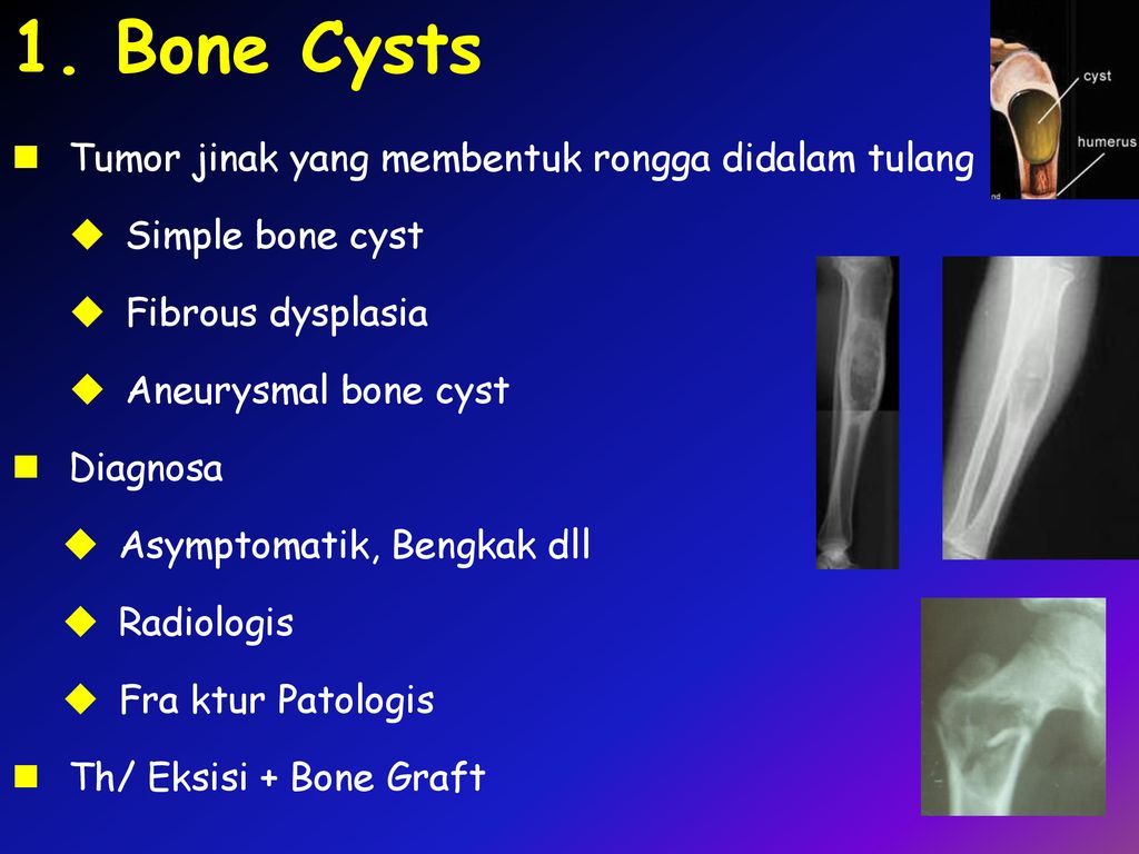 No treatment is needed, but these can be surgically removed if they are bothersome.
Sebaceous Gland Tumor
A sebaceous gland tumor is commonly found in older dogs. They are typically smaller than a pea and may develop in any location. Some will bleed or secrete a material that forms a crust. Large breeds often form these on their head, specifically their eyelids, and they may be black in color. Treatment is not necessary, but surgical removal may be considered when the growth is bothersome.
Meibomian
A meibomian gland tumor is a slow-growing benign lump that forms in the meibomian gland at the edge of the eyelid. The tumor can stick out or grow into the eyelid. They may become inflamed, irritated, painful, or ulcerated. They may also cause inflammation of the cornea and conjunctiva.
Large tumors may cause problems when your dog blinks, because they cause extra tearing and tear staining. They are diagnosed by their appearance and location, and they can be removed with surgery, or the tissue can be frozen for removal.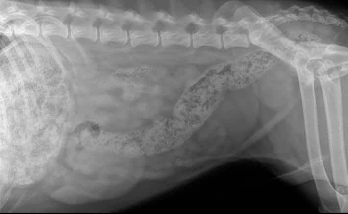 They rarely grow back after removal, but regrowth is possible.
Epulis
An epulis is a common benign growth found in the mouth of dogs. They may form when a tooth rubs against the gums, as with an underbite in brachycephalic (flat-faced) breeds.
These smooth, fleshy, pink bumps grow on the gum tissue near the outer surface of an incisor, canine, or premolar tooth. They can appear to grow on a stalk of tissue, like a mushroom, or as an unmoving mass, and they may have a bony interior. Certain types can invade surrounding bony tissue.
Diagnosis of an epulis is made by recognizing it from appearance and confirming with a biopsy. An x-ray of your dog’s head will show if it has invaded surrounding tissues. These growths should be removed surgically, along with the adjacent tooth and any bony tissue that may be affected. They do not usually regrow if the entire tumor is removed. Radiation therapy may help cases where the growth is inoperable.
Follicular Cysts
Follicular cysts are large, benign bumps on the skin that grow up from the hair follicle. They may release a thick material that is white, yellow, or brown when you push on them. As they get bigger, they can become itchy or painful.
These types of cysts are diagnosed on physical examination and may be confirmed with microscopic examination of a small sample of cells aspirated with a needle. Follicular cysts may become infected and require antibiotic treatment. If they are growing or become painful, they may be surgically removed and should not regrow.
Perianal Adenomas
Perianal adenomas are benign growths common in older, unneutered male dogs. They grow from oil glands near the anus but can also occur in similar glands along the abdomen, on the back, and near the tail.
These commonly show up as multiple small lumps. Larger tumors may develop bleeding ulcerations and can compress the anal canal, making it difficult for your dog to poop.
Almost all male dogs are cured by castration alone, but large or ulcerated tumors may also be surgically removed. Females improve with surgical removal, but the growths often recur. Laser surgery or freezing the growth may be necessary to avoid fecal incontinence when surgery involves the anal sphincter.
Hemangiomas
Hemangiomas are benign tumors that occur in adult dogs and closely resemble blood vessels. You usually see them on a dog’s legs and trunk. They may be single growths or multiple, compressible, reddish-black circular lumps that can resemble a blood blister. Some may become large and even ulcerate. The recommended treatment is surgical removal.
Nevus
A nevus is a dark raised or flat benign growth on the skin, commonly called a mole. These are found on areas prone to trauma, such as the legs, head, and neck, usually in older dogs. Treatment is surgical removal.
Trichoepitheliomas
Trichoepitheliomas are small, benign lumps that pop up from the hair follicles of adult dogs. They are cyst-like and filled with condensed, yellow, cheesy, granular material. They can occur anywhere on the body, but especially on the face and trunk. Treatment is surgical removal, but they are likely to continue to form at other locations, even after surgery.
They can occur anywhere on the body, but especially on the face and trunk. Treatment is surgical removal, but they are likely to continue to form at other locations, even after surgery.
Cornifying epitheliomas
Cornifying epitheliomas are benign growths that stick up from the skin surface and look like horns. They arise from hair follicles and may form anywhere on a dog’s body, but they are more common on the back, tail, and legs of adult dogs. No treatment is necessary unless there is evidence of self-trauma, ulceration, or secondary infection. Surgical removal is the ideal treatment.
Basal Cell Tumors
Basal cell tumors are benign growths that develop on the head, ears, neck, and forelimbs of older dogs. They are raised swellings that are typically firm, solitary, dome-shaped, and small. Some may be hairless, ulcerated, and stick out like stalks from the surface of the skin. They are dark in color and may form cysts that break open and drain fluid or pus. Treatment is surgical removal, especially when the dog is uncomfortable.
Malignant Tumors
Malignant tumors are cancerous growths that can invade tissue and spread to organs.
Angiosarcomas
Angiosarcomas are highly malignant blood vessel tumors that may vary in appearance. One or more red lumps in the skin or underlying soft tissue are commonly seen, but they may also appear as a poorly defined bruise.
All types grow rapidly and destroy surrounding tissue. They also spread to the lungs and liver. Angiosarcomas may occur in response to sun exposure in dog breeds with short, white coats, but dogs with dark, thick coats can develop them as well.
They usually form on the underside of the trunk, hip, thigh, and lower legs. A biopsy is required for a diagnosis. Freezing and laser surgery can help control smaller surface tumors. Surgical removal is needed for tumors below the skin’s surface. Chemotherapy may also be recommended to treat any remaining tumor cells.
Basal Cell Carcinomas
Basal cell carcinomas are flattened or raised growths that appear anywhere on the body of an older dog. They may spread to surrounding skin, forming new ulcerations, but they rarely spread to other organs. Surgical removal is recommended, including enough skin around the tumor to ensure that no tumor cells remain.
Liposarcomas
Liposarcomas are rare but may develop in older male dogs on the chest and legs. They can be soft or firm lumps that are slow to spread to other locations. Treatment is surgical removal, but recurrence is common. If this happens, radiation treatment may also be required.
Lymphosarcoma
Lymphosarcoma rarely develops directly on a dog’s skin but may be seen as a surface tumor or along with internal tumors. It can look like flaky skin, red patches, raised and ulcerated areas, or lumps deep within the skin.
There are two forms of skin lymphosarcoma that differ in their expected progression and response to treatment, so it is important to determine which type your dog has early on. Treatments include surgical removal, chemotherapy, and radiation, done separately or combined. These treatments may improve the signs of the disease but do not lengthen the dog’s life expectancy.
Mast Cell Tumors
Mast cell tumors are the most common malignant tumor seen in dogs. They often affect older dogs but can occur in dogs of any age.
They develop solitary growths anywhere on the body, especially the limbs, lower abdomen, and chest. Larger or rapidly growing tumors and those in certain locations are more likely to spread. Their appearance can greatly vary, but most are raised and either soft or firm to the touch.
Your vet will need to examine a sample of cells from the growth under a microscope to confirm a diagnosis. There is variation on how aggressive these tumors are. Surgical removal is necessary. If the tumor regrows or spreads, other treatments, including chemotherapy and radiation, may be used.
Malignant Melanomas
Malignant melanomas are another type of skin tumor of older dogs. They commonly develop on the lips, mouth, and nail beds of male dogs. They appear as raised, ulcerated lumps and may be dark, light gray, or pink. If they appear in the nail bed, the toe is often swollen.
These tumors grow quickly and may spread quickly to other organs. Complete surgical removal is the preferred treatment, but it may be difficult and involves removal of adjacent tissue to prevent recurrence. Chemotherapy and radiation are not effective treatment options. A vaccine is available that helps shrink the size of the tumor, which may prolong your dog’s life expectancy.
Fibrosarcomas
Fibrosarcomas are common, fast-growing malignant tumors in dogs. Most are on the trunk and legs and vary in appearance and size. Those under the skin’s surface appear lumpy, while those deep under the skin may be firm and fleshy.
They can invade underlying muscles, but most do not spread to other areas of the body. Treatment consists of surgical removal, though complete removal may not be possible, and regrowth is common. Fibrosarcomas may also be treated with radiation and chemotherapy.
Squamous Cell Carcinomas
Squamous cell carcinomas can be found in two places on a dog: on the surface of the skin or under a nail. Most appear as firm, raised, irregular, and ulcerated areas. Many are solitary, but areas of prolonged sun exposure may produce multiple tumors. These growths invade surrounding tissues. Some are slow to spread, while others grow more rapidly. Treatment involves complete surgical removal of the tumor along with some normal tissue.
What to Do If You Find a Lump or Bump on Your Dog
When you find a growth on your dog, have your vet do a physical exam. It’s helpful to note the location of the lesion, how long it’s been there, any changes that have occurred since you first noticed it, and whether your dog seems bothered by the growth.
How Vets Diagnose Lumps, Bumps, and Cysts on Dogs
A sample of cells may need to be taken and evaluated under a microscope for a diagnosis. This can come from taking an impression of the surface of the growth, using a syringe and small needle to withdraw a small sample of cells in the exam room (fine needle aspiration), or surgically removing a small tissue sample (biopsy) while your dog is under local or general anesthesia.
Most veterinarians evaluate impression smears or fine needle aspirates by staining the slide and examining it under a microscope in the veterinary office. Trained veterinary pathologists are available to analyze these same samples or small tissue samples to determine a diagnosis. Then your vet can determine the appropriate treatment recommendations and explain the expected outcome.
Treatment for Dog Lumps, Bumps, and Cysts on Dogs
Options for treatment of a growth on a dog may include:
-
Monitoring for changes
-
Removal by freezing or laser treatments
-
Surgical removal of the lump with or without also removing some normal tissue
-
Chemotherapy
-
Radiation
How to Monitor Your Dog’s Lumps and Bumps
Keep a log where you write down when you first noticed the lumps and/or bumps, how many there are and where they are located, the size, color, and texture, whether it’s moveable or seems to be fixed to underlying tissue, and whether there is any discharge present.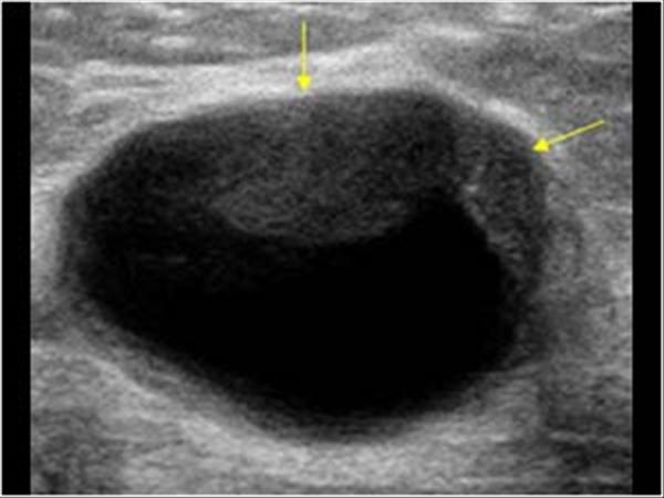 Take pictures and note any changes from day to day in any of these factors.
Make an appointment with your vet as soon as possible and bring your log and photos along with any questions you may have.
FAQs
What does a sebaceous cyst on a dog look like?
A sebaceous cyst is a smooth, raised swelling that contains sebum from the oil glands associated with hair follicles. They can become infected and inflamed and may rupture, secreting a thin, yellowish discharge.
What can you do for a dog with a sebaceous cyst?
A sebaceous cyst should be observed for changes in size or material within the cyst. The cyst may break open from being scratched and release a fluid-like material. This should be gently cleaned with warm water and then allowed to dry. Any sign of infection or sensitivity in the area should be evaluated by a veterinarian for treatment recommendations and possibly surgical removal.
How do I get rid of bumps on my dog?
The safest way to remove bumps from a dog’s skin is to have a veterinarian diagnose the type of bump.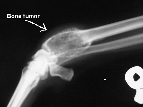 Then they will determine an appropriate plan for surgical removal using local or general anesthesia.
Can a bug bite cause a bump on a dog?
Yes, a bug bite may cause a temporary swelling on a dog. These may be itchy as well. Growths are usually more defined and rarely regress but remain the same or grow larger over time.
Is a belly lump normal after a dog spay?
No. A belly lump may indicate a reaction to a buried suture, or rarely, a hernia of the abdominal wall. Contact your vet if your dog has a lump in their belly after being spayed.
Should I have my dog’s lump removed?
This would depend on the type of lump that it is. Many benign growths do not need to be removed. However, if the growth changes in appearance, bothers the dog, or interferes with movement, then a veterinary exam is needed to determine the appropriate treatment. For malignant growths, your vet can determine the appropriate treatment.
Featured Image: iStock.com/Elayne Massaini
Treatment of breast tumors in domestic animals
What is a tumor?
An overgrowth of abnormal tissue that looks like normal tissue in the body.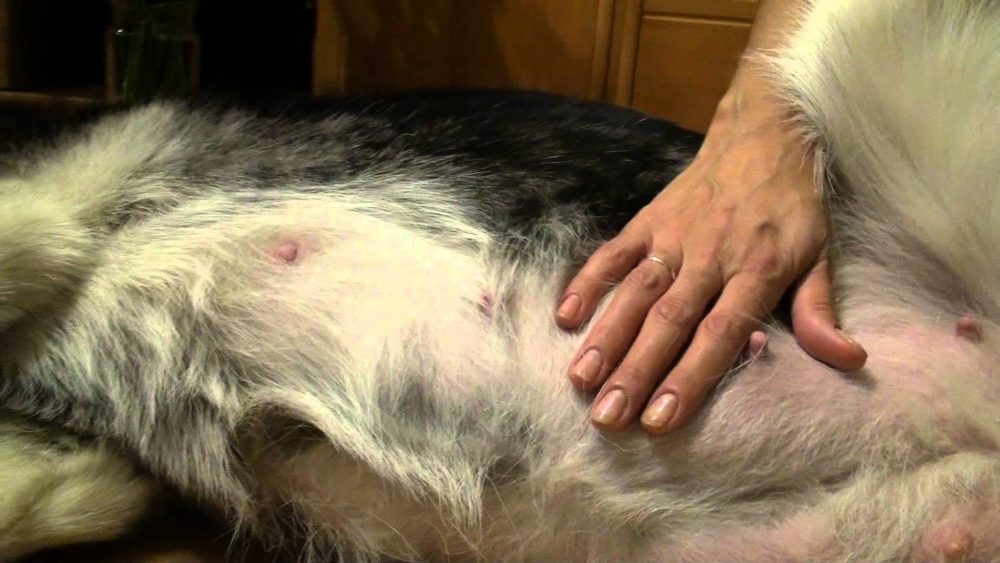 Tumors or neoplasias or neoplasms can grow from any tissue.
How often do you encounter breast tumors in animals?
20% of all animals who applied to the clinic are animals with neoplasms, most often with tumors of the mammary gland (MT), less often – tumors of the spleen, liver, skin, bones.
Which animals are more likely to have AMF, at what age?
More common in cats over 4-5 years of age, in non-neutered cats, and in those whose owners used hormonal drugs to suppress estrus. In dogs – females older than 5 years, not sterilized, but most often their tumors are not associated with the use of sex hormones, they are given to dogs extremely rarely.
Are there stages in breast tumors?
Of course, there are, like any other tumors. The stage that the doctor will put at the appointment will depend on the size of the tumors, the involvement of regional lymph nodes in the process, and the presence of distant metastases.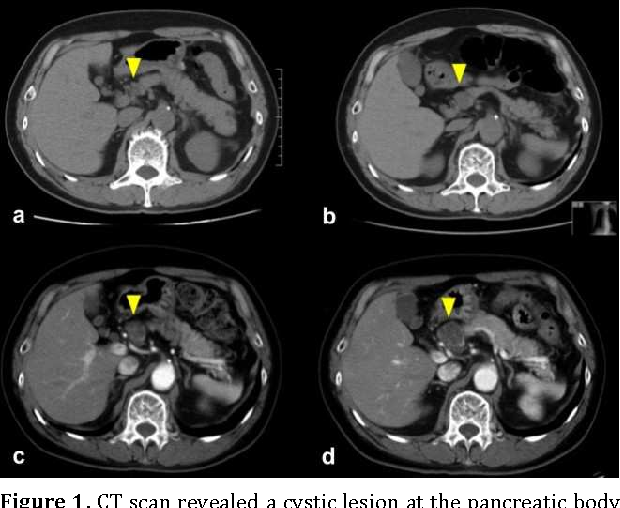
What examinations should be performed on the animal before the operation?
It is obligatory to take general and biochemical blood tests, ultrasound of the heart, ultrasound of the abdominal organs and chest X-ray before the operation. It is also necessary to do a histological examination of the tumor after the operation. Blood tests are needed to know how the body works in general, whether there is kidney failure. An ultrasound of the heart of a dog or cat is necessary to detect hidden congenital or acquired pathologies. Ultrasound of the abdominal organs – to detect tumors or metastases in the internal organs. X-ray for animals in the chest area – to exclude metastases to the lungs. If there are distant metastases on these studies, the operation is not advisable. If there are no metastases, everything is fine according to analyzes and other studies, then it is necessary to tune in to surgery with subsequent examination of the tumor.
Is surgery always necessary?
Yes, always.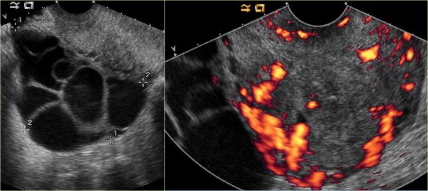 The operation is performed so that the tumor does not increase and does not grow. Because the tumor tends to mature and open like an abscess. Even if the owners do not agree to the operation due to the fact that the tumor is small and has been in one place for more than one year, it still needs to be removed. Such a seemingly harmless tumor can metastasize to the lungs, and changes will be noticeable only in the last stages. A cough will appear, or the animal will refuse to eat, and only on an ultrasound or x-ray can one detect the presence of metastases.
When is chemotherapy administered to a dog or cat?
Chemotherapy is carried out only after the results of histological examination. It is advisable to do this always if the tumor turned out to be malignant. Even after a successful operation, tumors can grow in the same place. Chemotherapy will not give a full recovery, but with the help of these drugs we will be able to stop the process of further appearance of malignant cells.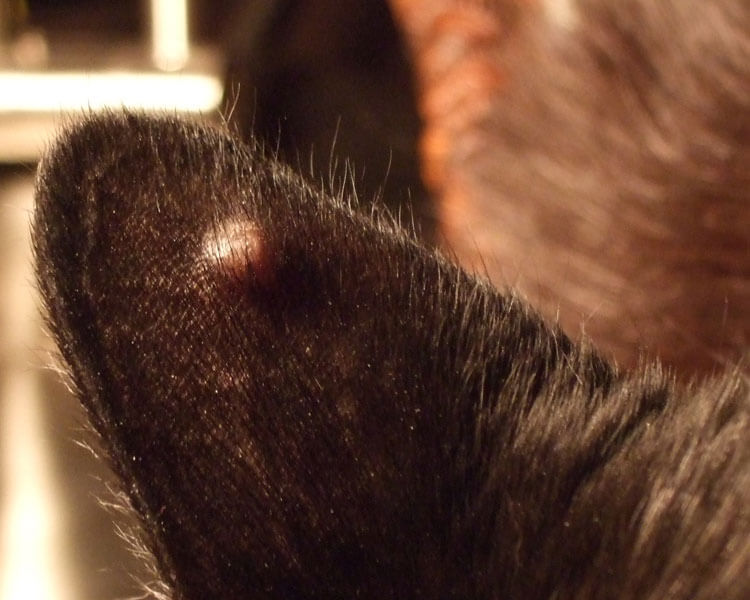
Does spaying of cats and dogs affect the development of AMF?
Cats and dogs spayed before their first estrus have a 70% reduced risk of developing mammary tumors compared to non-spayed cats. If a cat or dog was neutered after the first estrus, the risk is reduced by 30%, if after the second, the risk remains the same for neutered and non-neutered.
What to do if a breast tumor has opened?
It is necessary to come to the clinic so that the doctor can assess the nature and localization of the wound, its extent. It is also necessary to assess the condition of the animal in order to decide with the owner whether it is possible to operate the animal in this condition. If not, then the doctor will prescribe antibiotic ointments or antibiotics for systemic use.
What is inoperable AMF?
The tumor is large and there is not enough healthy tissue to close the wound after surgery. An extensive tumor that has grown into muscle tissue, bone tissue, and if there is damage to internal organs.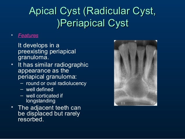
What determines the operation area?
From the location of the primary tumor. In cats, regardless of location, all milk bags on one side are removed. In dogs, the location of the tumor plays a role. sometimes it is possible to remove half. The area of the operation can be assessed by the doctor at the reception. If the cat does not have neoplasm nodules on the second side of the mammary gland, in order to improve the quality of life and health of the patient, it is still recommended to remove the second side of the mammary gland packets one month after the first operation. In dogs, most often half of the packages with neoplasms are removed.
What is a mastectomy? What is it like?
Mastectomy – removal of the breast. When performing a local mastectomy, the tumor itself is removed directly. This is done only if the animal is old, mainly such operations are performed to improve the quality of life of the animal, and to be able to take material for histological examination.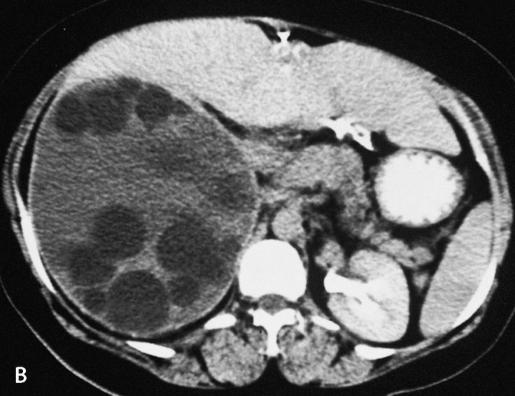
Regional mastectomy is only possible in dogs. Remove 3 milk packs out of 5 on one side. Unilateral mastectomy removes one side of the breast, bilateral mastectomy removes both sides
Is it necessary to sterilize the animal together with the removal of AMF?
Optional. Except for the presence of a cyst or neoplasm of the ovary and with inflammatory processes in the uterus.
Do males have AMF?
Very rare. The treatment is the same as for self – removal of tumors with subsequent research.
What is the prognosis after AMF removal?
The prognosis will depend on the type of tumor. If the tumor is benign, the prognosis is good. If malignant, the prognosis is doubtful or unfavorable. In the presence of a malignant tumor, we can prolong the life of an animal for a short period of time – from 3 months to 2-3 years. However, we have cases where animals lived 6-7 years after the operation. For an animal, this is almost half of life.
For example, a cat is 16 years old. Is it possible to have surgery at this age?
Possible. In our clinic, several dozen operations were performed on cats over 15 years old, 6 operations on cats aged 20-23 years. All operations ended favorably, we extended the life of the animal by an average of 2 years.
What postoperative complications can develop?
During the removal of the mammary gland, a large amount of tissue is removed, and such wounds heal longer than from castration or spaying of dogs and cats. Most often, the owners do not fully understand the type of operation. The occurrence of extensive hematomas (which is the norm) is perceived as a critical situation. Also, postoperative seroma – the accumulation of fluid in the groin area is also the norm.
What tests should an animal undergo after AMF removal?
Obligatory every 3-6 months to do ultrasound of the abdominal organs, chest X-ray for early detection of metastases. In the presence of metastases, we will not be able to help the animal, however, the owners will be mentally prepared for an unfavorable outcome.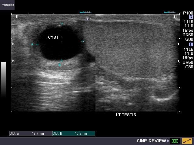
Is a histological examination always necessary?
This study will help us to identify whether the tumor is benign or malignant. If it is malignant, then we need to know what type of tumor it is so that we can select drugs for chemotherapy.
The owners took a biopsy to remove the tumor. The tumor turned out to be benign. Should it be removed or can it be left on?
It is better to remove such a tumor, because benign tumors can also continue to grow, or become malignant after some time. This moment of transition cannot be tracked by any analysis other than a biopsy.
What is palliative surgery?
These are operations that are aimed at improving the quality of life of the animal, but do not have any therapeutic effect. For example, we can remove an open tumor from an old animal with metastases. This tumor will not cure the animal, but it will make life much easier for both the animal and the owners.
Veterinary surgeon Alexander Stanislavovich Vasilkin
Castration of dogs (male) in Minsk in the AibiVET veterinary clinic.
Canine brain tumor: treatment and care
Published in
Private Oncology
Author
Mikhail Shelyakov
May 14 2018
Rate the material
- 1
- 2
- 3
- 4
- 5
(2 votes)
Brain tumors occupy only a small percentage of the total number of neoplasms.
But at the same time, they are distinguished by the extreme severity of the symptoms and the direct threat to life.
There is a big diagnostic problem. Clinical signs that allow suspecting a tumor process are mainly due to the symptoms of increasing intracranial pressure.
In the initial stages, these may be short-term epileptiform seizures, then they become more frequent and more severe.
With further growth of the tumor, there is a change in consciousness, prolonged erratic movements, convulsive tension.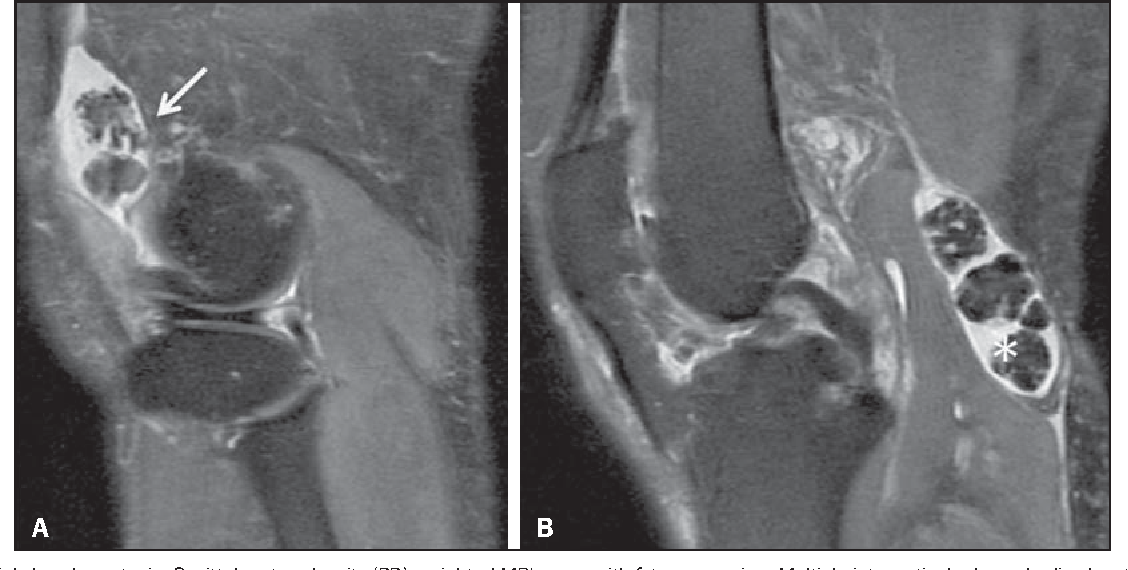
Alarms – blurred vision, falls, loss of consciousness, chaotic eye movement.
The leading diagnostic method for suspected brain tumors in cats and dogs is MRI, but doctors observing the patient do not always understand the severity of the animal’s condition, delaying the study. It can be difficult for an inexperienced doctor to suspect a dog’s tumor in the head, even with obvious symptoms. Therefore, animals usually come to us in poor condition, having undergone more than one course of ineffective treatment. As a result, we often have to go for broke, limiting ourselves to a diagnostic minimum that allows us to establish a brain tumor and its exact localization. And try to remove it surgically if technically possible. And here comes the second big problem.
The second big problem. Technical difficulties of the operation. In order to remove a diagnosed brain tumor, we perform a craniotomy to access the brain. The size of the trepanation window depends on the volume and location of the neoplasm.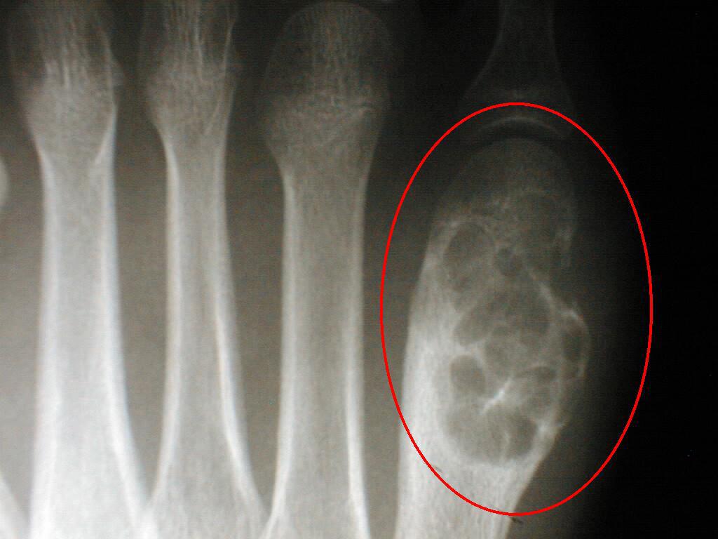 Through the window, the tumor is removed (if possible) and intracranial decompression is provided.
In the vast majority of cases, meningiomas are found. These are benign tumors located outside the brain. They can be removed completely. The difficulty lies in the fact that it can be difficult to visually determine the boundaries of the neoplasm in situations of restricted access.
Intraoperative ultrasound can be performed to assess the morphological state of the brain. Moreover, if the operation is successful, the trepanation window does not close immediately. For some time, the state of tissues is monitored through it. As a rule, if re-growth is not detected within three months and the patient’s condition is satisfactory, then an additional operation is performed to close the skull defect. Usually the trepanation window is closed by its own bone lid. She, before the second operation, is sewn under the skin next to the surgical wound. The method of preserving one’s own bone fragments in this way is called “biopreservation”.
Trepan window. Wide access. The tumor was diagnosed by MRI with contrast. Operation stage.
There are situations when a tumor on the head is visible from the outside, as a dense bone painless formation. It does not necessarily mean that it is a brain tumor. Most often, these are third-party tumors that grow in all directions, while deforming the bones of the skull. The latter “pressing” into the cranium, in turn, create compression of the brain with all the symptoms similar to those of a true brain tumor.
Such situations also require surgery – removal of the tumor and decompression of the brain.
There are cases when not a tumor, but a cyst forms in the brain. It’s like a vial of liquid. And this cyst occupies the volume and puts pressure on the brain, akin to a neoplasm and can cause all the same symptoms that make it possible to suspect a brain tumor.
But a cyst is removed much easier than a tumor. It is only necessary to carefully determine its coordinates and find out how accessible it is for a surgical operation.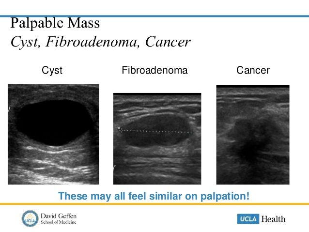 If everything is done and everything is available, then a small trepanation window is formed in the bones of the skull and the cyst is removed through it. The window can be of any shape. The choice of configuration depends on the tasks. But its dimensions are usually insignificant, which allows not to close the bone defect after the operation, as is done after a wide trepanation.
Our personal statistics: satisfactory outcome after removal of brain tumors in about 10% of cases (good quality of life, survival over a year). The third part dies during the operation or immediately after it. In another third of the operated animals, the lost neurological deficit is not restored. Or, in other words, the operation does not bring results. Everything remains as it was.
The statistics could be improved many times over. But, as mentioned above, almost all animals come to us deeply healed and in poor condition.
We wish you to avoid terrible diseases. But if a misfortune has already happened, we will apply all our experience and strength.
Doctor of Veterinary Medicine M. Shelyakov
Mikhail Shelyakov
Latest post from Mikhail Shelyakov
- Lipoma in cats
- Tumor of the spleen in cats
- Cancer in cats
- Dental care for a dog
- Paw pad disease in dogs
More in this category:
« Sparing organ-preserving operations
Wen in dogs: causes of lipoma in dogs, treatment, photos
Top
Bumps, cysts and growths in dogs
This article will tell you:
And now more.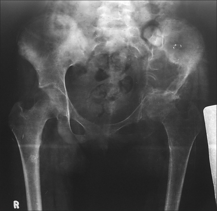 ..
..
Dogs often have growths, cysts and bumps. They can appear in dogs at any age, but they are one of the most common health problems seen in older dogs.
As a dog owner, it’s good to know what growths you may encounter. Any persistent, unusual bump, cyst, or growth requires a visit to the veterinarian.
01. What are bumps, cysts and growths in dogs?
Most veterinarians will refer to any unknown lump or tumor as a growth, cyst, or mass. In general, these terms can be used interchangeably, but most veterinarians avoid the word “tumor” unless the tumor is defined as a type of cancer.
02. Symptoms of Tumors, Neoplasms and Cysts in Dogs
Abnormal growths can occur anywhere on the body or in the mouth.
Warning signs include:
- Abnormal or very small to very large lump
- Discolored, itchy or irritated skin over the lump
- Swollen area (especially inside the body)
- Enlarged lymph nodes
- Lameness or tumor affecting bone
03.
 Sebaceous cysts, adenomas and adenocarcinomas
Sebaceous cysts are common types of skin cysts that contain sebum, a thick oily material commonly found in the skin around hair follicles.
These growths can be anywhere on the body. Sebaceous cysts are benign, but they can also be mistaken for a malignant tumor called sebaceous adenocarcinoma or a benign growth called sebaceous adenoma.
If your dog is not bothered by the cyst, the veterinarian may leave it alone, but the cyst can be surgically removed if necessary.
Once removed, the cyst must be sent to a laboratory so that a veterinary pathologist can determine that it is indeed just a sebaceous cyst, adenoma, or adenocarcinoma, which may require additional treatment.
04. Moles on the skin
Moles on the skin of dogs are similar to those on humans. Some can become quite large and saggy, dangling from the skin on a narrow stalk.
Moles on the skin are benign and are not usually removed unless they bother the dog or become very large and irritated.
05. Histiocytomas
Histiocytomas are red bumps that can appear quickly on your dog’s skin and usually go away on their own within a few months. Although these are benign tumors, some of them can grow quickly and really bother your dog.
Your veterinarian may recommend removal of large or irritated histiocytes. Unlike other common skin lesions, histiocytomas are most commonly diagnosed in young dogs.
06. Squamous cell carcinoma
Squamous cell carcinoma is a type of tumor that can be caused by sun exposure. This type of cancer can occur on the skin and/or in the mouth. These tumors may be pink or reddish in color and have a misshapen, “raw” appearance.
07. Malignant melanoma
Malignant melanoma can occur on the skin and/or in the mouth and is thought to be caused by sun exposure. Many of these tumors are black, but not all of them will look the same.
08. Mouth Growth in Dogs
Your dog’s mouth may develop a variety of growths.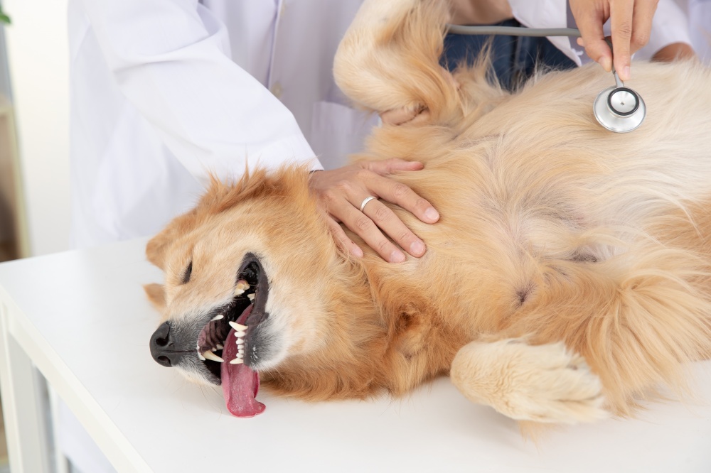 Some growths are difficult to see but cause symptoms such as bad breath, chewing problems, difficulty holding objects in the mouth, pain in the mouth, and scratching on the face or mouth. Of course, these signs can also indicate dental disease and should not be ignored.
Papillomas are warts caused by the papilloma virus. They can appear on the lips, face, and mouth of the dog. Papillomas are benign but highly contagious. They can be removed if they are causing problems for your dog, but in many cases they resolve on their own.
Epulis is an oral growth that usually forms on the gum tissue around the tooth. Many epulides are benign, but some may be malignant, so further diagnosis is needed.
Gingival hyperplasia is a benign growth of gum tissue that may look a bit like a tumor in some dogs. This extra gum tissue can be removed if it affects the teeth or bothers the dog. The removed tissue may be sent to a veterinary pathologist to ensure there are no cancer cells.
Oral melanoma may occur in the mouth and may be black.
Squamous cell carcinoma and fibrosarcoma are other common types of cancer that can develop in the oral cavity of dogs.
Some oral tumors can affect the teeth and bones in the mouth and face. If your dog has a mouth mass, your veterinarian will likely recommend placing it under anesthesia so that a thorough examination and x-rays can be taken.
09. Lipomas in dogs
Lipomas are a common type of tumor in dogs. A lipoma is a benign fatty mass that can be found anywhere on a dog’s body, usually under the skin. They are usually soft and movable and rarely cause pain or discomfort to the dog.
Lipomas can be surgically removed if they interfere with your dog’s mobility or comfort, grow rapidly, or rupture (causing skin damage). In rare cases, an apparent lipoma is actually a malignant tumor called a liposarcoma. Diagnostic testing can tell them apart.
10. Fat cell tumors in dogs
Fat cell tumors can occur as skin bumps or internal tumors.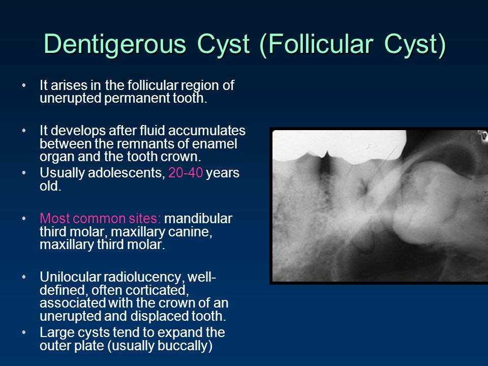 These growths can release histamine if disturbed, which can have a negative effect on your dog’s body.
If your veterinarian suspects a fat cell tumor, your dog may first be treated with diphenhydramine to minimize histamine release.
After removal of the mass, the pathologist grades the tumor as I, II, or II. This score tells you how cancerous the tumor is and how likely it is to metastasize (spread to other parts of your dog’s body).
11. Mammary tumors in dogs
Breast tumors are more common in bitches, especially those that are not neutered, but can occasionally occur in neutered bitches.
Although some breast masses may be benign, many are malignant. The prognosis improves when the growths are diagnosed and surgically removed when they are small.
Breast cancer in dogs
12. Internal tumors
Some dogs develop internal masses in the chest or abdomen, especially older dogs. Internal growths may be discovered because of the symptoms they cause (such as difficulty breathing or vomiting) or during a routine physical examination.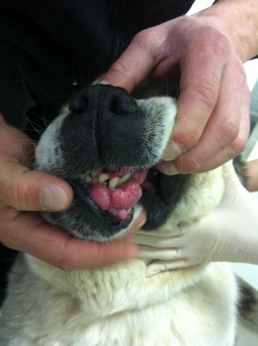
Internal masses can be benign or malignant and are usually definitively diagnosed by a combination of radiographs, ultrasound, laboratory tests, and biopsy. Treatment depends on the location and type of tumor.
Lymphoma is not actually a tumor; it is a cancer of certain cells of the immune system. However, the first sign of lymphoma is often swollen lymph nodes, which may look and feel like tumors. 4
Pet owners most commonly notice lumps in the neck, but they can also be found in the armpits (armpits), groin (lower abdomen near the thighs) and the back of the knees. Lymphoma is often diagnosed by fine needle aspiration or biopsy. Chemotherapy is the most common treatment for lymphoma.
14. Causes of tumors, neoplasms and cysts
- Sebaceous cysts, adenomas and adenocarcinomas
- Moles on the skin
- Histiocytomas
- Squamous cell carcinoma
- Malignant melanoma
- Canine oral growth
- Lipomas
- Mast cell tumors
- Breast tumors
- Abdominal weights
- Canine lymphoma
- . .. and more, including cysts (not sebaceous glands), hematomas, infections, inflammatory reactions, and tumors resulting from trauma.
 .. and more, including cysts (not sebaceous glands), hematomas, infections, inflammatory reactions, and tumors resulting from trauma.
 15. Diagnostic process
When a tumor is found, your veterinarian will perform a physical examination. If the tumor is very new and potentially temporary (for example, the result of an insect bite or injection), a veterinarian may recommend an observation period, but in most cases, they will perform additional diagnostics to determine the type of cells that make up the mass.
This usually means taking a sample of material from the mass and analyzing it under a microscope.
The veterinarian will usually collect these samples by fine needle aspiration or biopsy. Evaluation of the specimens (often performed by a pathologist) can indicate whether the growth is malignant and, if so, what type of cancer is present.
If your veterinarian diagnoses your dog with cancer, additional diagnostics will likely be recommended, including:
- Laboratory tests such as a blood chemistry test, complete blood count, and urinalysis.
- Radiographs (x-rays), which may show signs of metastases or other problems.
- Ultrasound, which allows you to better view the internal organs and identify metastases.
- CT scan or MRI to help veterinarians better understand the structure of your dog’s tumor and some internal organs.
Some advanced diagnostics and treatments must be performed by a veterinarian.
16. When your dog needs a biopsy
If fine needle aspiration is not effective (or if the veterinarian believes it is not the best option), a biopsy is usually the next recommendation.
Biopsy is often performed on the dog under general anesthesia or sedation, but local anesthesia may be used instead depending on the size and location of the lesion.
The biopsy can be performed with a special large needle. Or the veterinarian may cut the mass surgically. In some cases, the entire mass is surgically removed and sent to a laboratory for identification.