


Save baby teeth stem cells: Should You Save Baby Teeth for Stem Cells?
The Tooth Bank – Frequently Asked Questions
The Tooth Bank – Frequently Asked Questions
What are Stem Cells?
Unlike other cells in the body, stem cells have the ability to transform into different types of cells and be used to regenerate tissue, bone, cartilage, and neural tissue.
What are Dental Stem Cells?
Dental stem cells are adult stem cells found in wisdom teeth and baby teeth. Dental stem cells are part of a group of adult stem cells know as “mesenchymal stem cells” and have the ability to differentiate into bone, dental tissue, cartilage, muscle, neural and other cell types. They are being studied for applications in regenerative medicine and dentistry.
What makes Dental Stem Cells different?
Dental Stem Cells are found in teeth. Primarily they are found in baby teeth and wisdom teeth. The stem cells that are located within teeth are called mesenchymal stem cells. These stem cells are used to regenerate bone and tissue throughout the body. There are currently over 2000 studies using Mesenchymal Stem Cells that are ongoing for treatments in regenerative medicine.
What diseases are being researched?
Currently there have been over 2000 clinical trials that have been done or are taking place regarding stem cells and regenerative medicine. There are a significant number of applications that are being studied using stem cells that include: Type 1 diabetes, Stroke, Parkinson’s, Alzheimer’s, muscular dystrophy, bone loss, multiple sclerosis, cardiovascular disease, neural injuries, and cancers (Leukemia, Lymphoma).
Why should I store dental stem cells?
There are several reasons; some have a family history or higher risk that prompts them to consider different options. Most see the future of stem cell research and don’t want to miss the opportunity to save their own stem cells.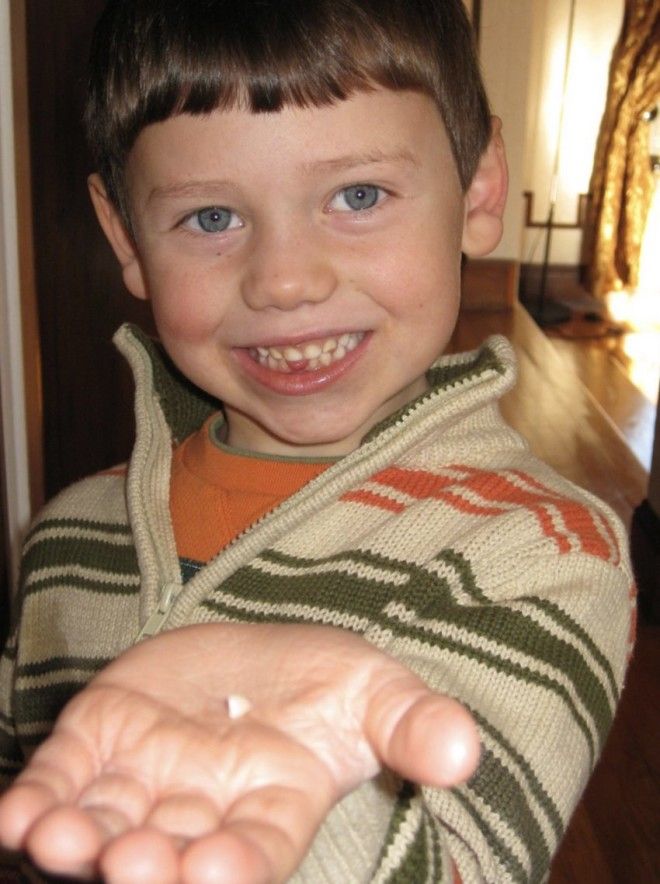
Should I bank more than one tooth?
Yes, banking more teeth will increase the number of cells. There is no additional fee to bank multiple teeth at the same time.
Why Should I choose Tooth Bank to work with?
When it comes to security, peace of mind, and affordability, there is no better option for you and your family. Tooth Bank provides first-class dental stem cell banking at an affordable price. Our team of scientists, dentists, and business healthcare professionals have decades of experience processing and preserving cells and are here to provide you with that peace of mind.
Multiple Children Banking
Yes, you should bank teeth from each child. Stem cells may not be a match between children.
Why cant I just pull out a tooth at the time its needed for cells vs. storing now?
Just like our bodies, teeth have more strength the younger they are and over time, like our bodies, enamel wears away and stem cells in teeth become less and less.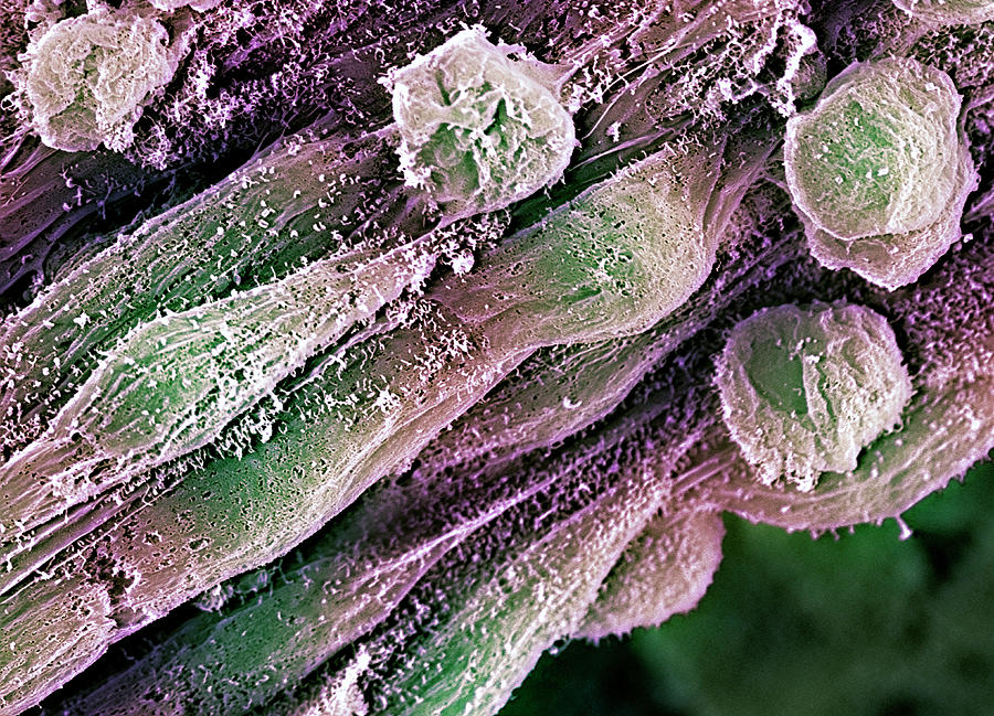 It is important to store now, when your children or family members are young so that the stem cells can be as healthy as possible.
What are the Tooth Bank Kit components?
In the kit you will receive all the necessary components for your dentist to collect:
- Collection Jar for teeth with solution already added
- Shipping container to return teeth in
- Pre-paid shipping label all completed
- Form for dentist to complete and return with the teeth
When should I enroll?
If you are expecting the loss of baby teeth or healthy tooth extraction in the next 365 days, you should order a kit today.
Do I need to let my dentist know I am planning on storing my teeth?
It’s best to let your dentist know that you are planning on storing the stem cells. Do let the dentist know that all is included in the kit.
Will my dentist know how to do it?
All dentists and oral surgeons have done this process. The Tooth Bank is always available to speak with your dentist at any time if they have any questions.
Can I store at home?
No, unfortunately, the cells need to be extracted from the dental pulp in the middle of the tooth, processed and then placed in cryopreservation. A freezer at home will not work.
Which teeth are best?
Wisdom teeth, baby teeth and healthy molars are best.
Do teeth with cavities work?
Some teeth with cavities work. It is dependent upon if the nerves and blood supply are still viable.
Do I need to bring anything to my dentists office?
All you need to bring is the collection kit.
Will my dentist charge me?
All the Dentist will be doing is placing the extracted teeth into the container. Fed Ex will pick up the kit at the dentist”s office, or you may drop off the kit at FedEX on the way home from the dentists office, or schedule FedEx to pick up at your home. There is no cost to you for Fed Ex to pick up your kit at home.
Can I pay for this with my flexible spending account?
Most Flexible Spending Accounts will let you pay for the annual storage fee but not the first years processing fee. It is best to check with your flexible spending representative.
Does insurance cover this service?
Unfortunately not. Storing your teeth is an elective procedure.
How long can stem cells be stored?
As of date, stem cells have been stored for over 22 years and have shown to be just as viable after 22 years as new samples.
How do I know my sample will be secure?
The Tooth Bank is FDA registered and HIPAA compliant, we follow all guidelines.
How do I insure my information is private?
The Tooth Bank is under HIPPA compliance rules, same as your dentist
What if my stem cells are not suitable for use?
This rarely happens, but in the event that in the processing your cells are found not to be viable, all monies you have paid will be refunded less $50.
Why saving your child’s baby teeth could be beneficial in the future
While parents have been saving their children’s baby teeth in memory boxes for decades, saving baby teeth for stem cells is a relatively new practice. Not quite as popular (or scientifically backed) as banking baby’s cord blood, storing baby teeth at a tissue bank may come with future benefits. That said, at this point, few experts are completely sold on the practice.
“The theory behind banking baby teeth is along the same lines of banking placental stem cells and umbilical cord blood — that the cells will be able to be harvested at some point to create other tissue,” says Dr. Amr Moursi, dental surgeon and professor and chair of the NYU Department of Pediatric Dentistry. “However, at this point, there’s not enough research and no FDA approved application — but perhaps 20 years down the road there will be, and your child will benefit.”
Wondering if you should hang on to your child’s baby teeth the modern way? Here, experts weigh in on keeping baby teeth for stem cells.
What exactly are stem cells?
Stem cells are cells with the potential to renew themselves into different types of cells within the body. In adults, they’re potentially found in tissues, such as bone marrow, fat and blood vessels. Other sources containing stem cells are three- to five-day-old embryos, as well as amniotic fluid and umbilical cord blood.
As the Mayo Clinic explains, stem cells are important, as they can generate healthy cells to replace diseased ones and increase the understanding of how diseases occur. By 2017, stem cell transplants benefited over a million people — including one woman whose body “woke up” two years after a stroke that left her severely impaired and another who became disease-free after battling Burkitt lymphoma.
As Dr. Giuseppe Intini, dental surgeon and associate professor of periodontics and preventive dentistry at the University of Pittsburgh and a faculty member of the McGowan Institute for Regenerative Medicine at the University at Pittsburgh, explains, regardless of their origin, stem cells are either totipotent, pluripotent or multipotent.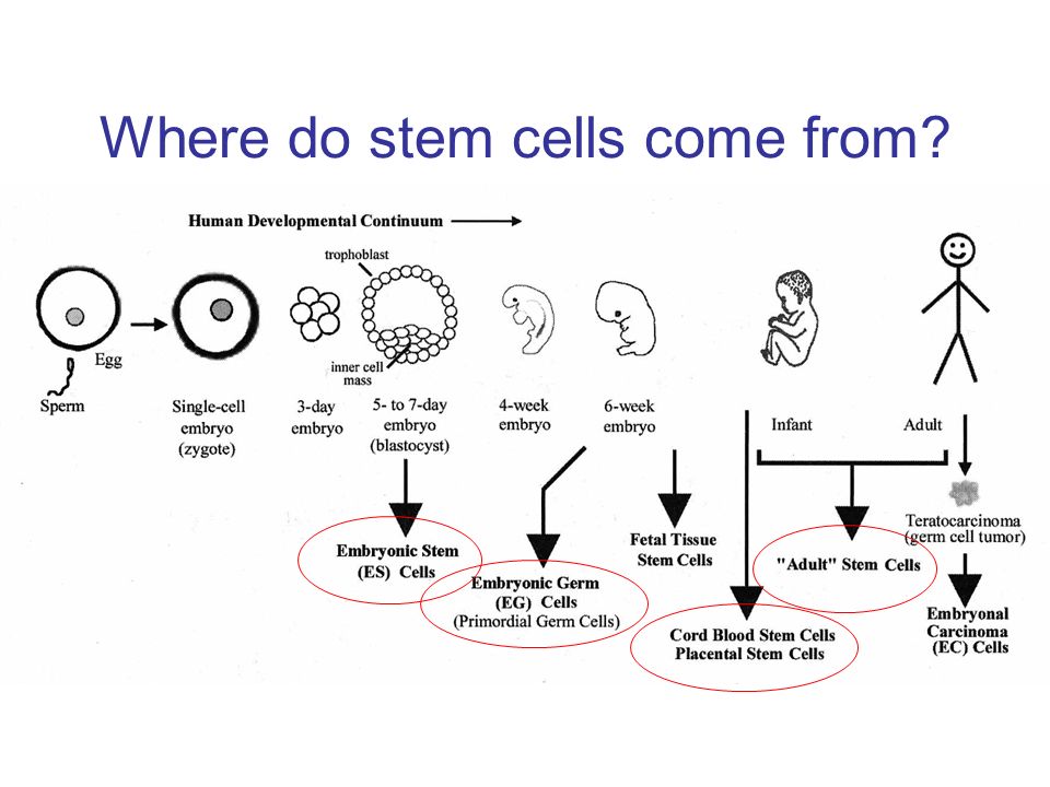
“Totipotent stem cells can generate any type of cell,” Intini says. “Pluripotent cells can generate all cells except the placenta, the amniotic sac and the umbilical cord (meaning a cell from the embryo can become anything from liver to hair cells but cannot fully generate another human being if transplanted in a host womb) and multipotent stem cells have the ability to develop into a limited type of cells. Teeth, it looks like, are multipotent.”
Can you use baby teeth for stem cells?
According to Intini, research is currently suggesting that there are stem cells in baby teeth — “suggesting” being key. “Right now, it appears — meaning, science is showing some evidence — that there are multipotent stem cells in teeth,” says Intini. “Keep in mind, though, this is all preclinical, and research has been done mostly with mice and rats. To really say teeth can produce stem cells that can be used for clinical application, we need clinical trials and right now there are very few. ”
”
It’s worth noting, though, that even though the clinical trials on stem cells in baby teeth are scant, they’re not altogether nonexistent. Research from a 2018 study suggests that “implantation of tooth stem cells can provide partial recovery of teeth injured by trauma.” Another found a connection between dental pulp stem cells and the treatment of mild to moderate knee osteoarthritis.
Should you keep baby teeth for stem cells?
With research being minimal at this point, and the high cost of properly storing teeth (more on that in a bit), neither Intini nor Moursi are completely sold on storing baby teeth at a tooth bank for stem cells.
“Right now, it seems that the stem cells in teeth can help repair teeth,” explains Moursi. “Frankly, dental regeneration [at the dentist’s office] is cheaper. If the research suggested stem cells in teeth could repair another ogan, like the heart or liver, that would be a different story.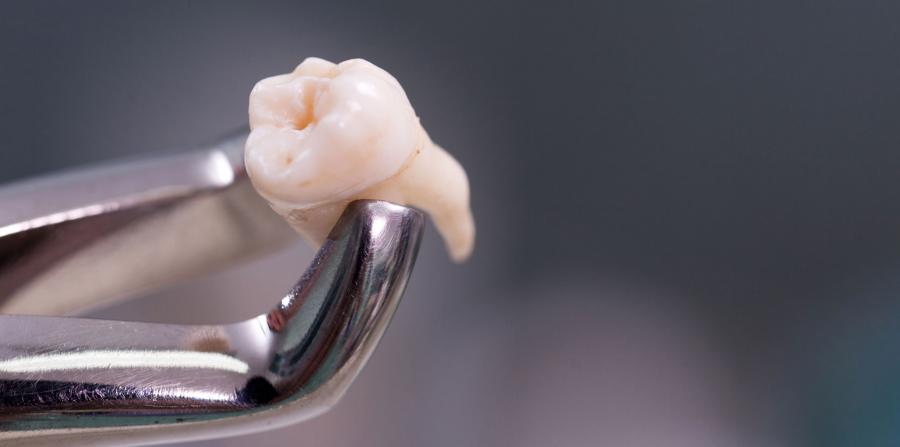 That said, saving baby teeth at a tissue bank — if you have the money — is an insurance policy of sorts and there’s no harm in doing so.”
How do you save baby teeth?
The old-fashioned way of saving baby teeth is to simply put them in a box (and, real talk, perhaps years later wonder why you kept them in the first place). But if you’re looking to save baby teeth for stem cells, the process is much different. (And, no, you can’t retrieve cells from teeth that have previously fallen out.)
Similar to the process of saving cord blood, baby teeth are saved in a kit that was previously purchased. “We send the kit to the client’s dentist and advise our clients to make an appointment with the dentist to have the tooth extracted when it’s a little loose,” says Art Greco, founder and CEO of the tooth tissue bank, StemSave. “Waiting for baby teeth to fall out on their own significantly reduces the chances of recovering healthy stem cells.” The likely reason being, Greco explains, that the blood supply to the pulp (where the stem cells reside) was terminated prior to the tooth falling out, “thereby rendering the cells dead.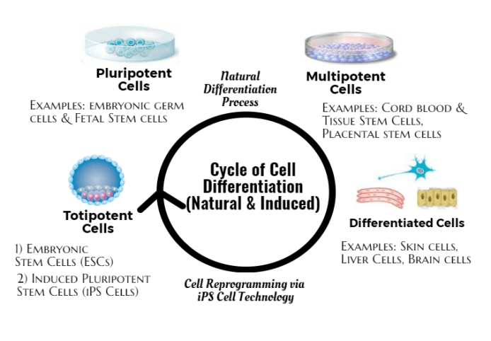 ”
”
After the tooth is extracted, the dentist places it in a kit designed to keep the cells alive during transportation. “We then arrange to have the kit picked up at the dentist’s office and overnighted to our lab for processing,” Greco says.
How much does it cost to store baby teeth?
Prices will vary at different banks, but there’s always an initial fee, as well as a monthly (or yearly) payment. At StemSave, there’s an initial recovery and processing fee of $630, as well as an annual storage fee of $120. Typically, payments are spread out in chunks, with StemSave offering three, six or 12 monthly payments.
“The cost is per specimen,” Greco says. “If a family sends us a kit with teeth in January and another kit in August, that would be considered two specimens and would incur two storage fees. However, if the family sends us a kit with more than one tooth — we process all the teeth in the kit and it is considered one specimen. We also have a number of payment options designed to accommodate families with a broad range of financial means.”
Also, it should be noted that, generally, insurance doesn’t pay for the cost of extracting a tooth at the dentist’s office solely for the purpose of preserving stem cells. “Insurance may cover the cost of the extraction if it is required for orthodontic reasons and it is a covered procedure,” Greco says. “However, in cases where the extraction is elective, insurance typically doesn’t cover it.”
Ultimately, keeping baby teeth in a bank is a personal — and financial — choice. Currently, there doesn’t appear to be tons of clinical evidence suggesting the stem cells found in teeth can do more than help other teeth, but with science constantly evolving, there’s no harm in doing so, if you have the means.
4 Reasons To Save Baby Teeth And Ways To Preserve Them
Saving baby teeth may help preserve childhood memories or even harvest stem cells.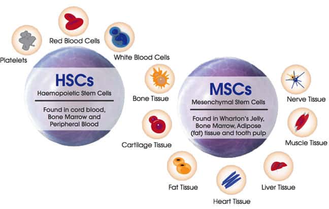
Research-backed
MomJunction believes in providing reliable, research-backed information to you. As per our strong editorial policy requirements, we base our health articles on references (citations) taken from authority sites, international journals, and research studies. However, if you find any incongruencies, feel free to write to us.
Image: iStockphoto
As a parent, you want to treasure every memory from your child’s development years. Hence, many parents save their child’s baby teeth. They may also do this to turn them into a memorable gift for the child when they grow older or to play out the child’s belief in the tooth fairy. Aside from these reasons, another incentive for keeping your child’s fallen teeth is also that they are a great source of stem cells (1).
Read this post to know why some parents save their children’s milk teeth, how to keep your children’s fallen baby teeth in good shape, what tooth preservation kits are, and how much it may cost to store baby teeth.
Why Do Parents Preserve Their Children’s Baby Teeth?
Here are a few possible reasons parents preserve their children’s teeth.
- Tooth fairy visit
As soon as a child’s milk tooth falls, it leads to excitement in children, as it’s time for the tooth fairy to arrive. Although a myth, little children find immense pleasure in keeping their fallen milk teeth under their pillow overnight in the hope of finding a gift they wanted in place of it in the morning.
- Keepsake for parents
Falling milk teeth is a sign that a child is growing up, which can be emotional for parents. Your child’s primary teeth can be a great keepsake for you for years to come.
- Gift for when the child turns older
A personalized item made from your child’s fallen and preserved milk teeth makes for a unique gift when they turn older. Imagine your child’s expression when they know their special gift was in the making for so many years.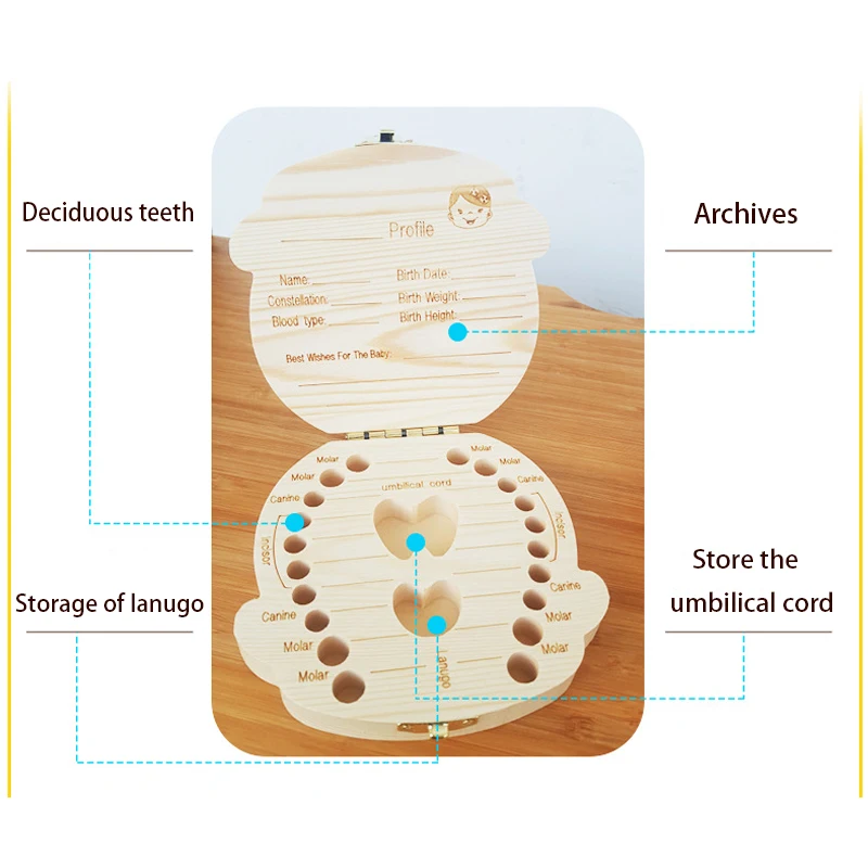 When your grown child is going through doubt that you ever cared about them, they will be touched to know you thought even their discarded teeth were worth saving. Maybe you do love them after all.
- Source of stem cells
Stem cells are found in tissues, such as the umbilical cord and the pulp of primary teeth and permanent teeth. These cells have remarkable regenerative properties that can help protect your child against many diseases and conditions in the future. Preserving your child’s baby teeth and banking them for these stem cells is highly recommended.
Stem cells have a remarkable potential for renewal. Due to this, they can give rise to different types of cells and tissues (2).
Recent research suggests that the pulp tissue is an excellent source of dental stem cells. These cells can be harvested from a child’s milk teeth as well as permanent teeth in children and adults (2).
What Are Some Ways To Preserve Your Baby’s Teeth?
If you wish to preserve your baby’s milk teeth for stem cell banking, contact your dentist or tooth bank as soon as your baby’s tooth falls out. The fallen milk tooth can be stored in cow or buffalo milk until it is collected by the tooth banking agency (3)
However, if you wish to preserve your baby’s teeth for their sentimental value or for playing along with your child’s tooth fairy fantasy, you can follow the following steps:
- Clean the teeth
Once your child’s baby tooth falls, clean it gently with soap and water.
- Disinfect
While cleaning with soap and water will remove the surface dirt, blood, and saliva, you might want to disinfect the tooth. This can be done by brushing the surface of the tooth with alcohol.
- Air-dry it
After cleaning and disinfecting the tooth, air-dry it. Drying the tooth prevents the growth of bacteria. You can use a dry cloth to wipe the tooth or place it in the Sun to remove the moisture.
What Are Tooth Preservation Kits?
To preserve your child’s teeth for stem cell banking, you will need to keep them in an appropriate kit. Although you can place the newly fallen teeth in a container of milk, tooth preservation kits are available for this purpose.
The American Dental Association (ADA) recommends that parents of young children keep an emergency tooth preservation kit handy. This kit consists of a container filled with sterile balanced salt solution (BSS), which is ideal for preserving your child’s fallen teeth (4).
This kit is also recommended in cases where a child’s tooth is accidentally knocked off. In these cases, you might want to visit your dentist within 30 minutes so that your dentist can try and fix the child’s tooth back in its socket (5). However, this is possible only with permanent teeth and not exfoliated milk teeth.
What Is The Cost To Store Baby Teeth?
Contact a tooth bank or tooth stem cell agency that collects and preserves babies’ fallen milk teeth. The average cost of collecting the tooth can range from $1,500 to $1,749, while the yearly cost of storing it for preservation averages around $120 (6).
Ideas For Preserving Your Baby’s First Tooth
Once you have safely cleaned and saved your baby’s milk tooth or teeth, you could try different ways to preserve them. Here are a few popular options you can try.
1. Keepsake box
The traditional practice of preserving baby teeth is followed by many parents across the globe. Thus, it is not difficult to find keepsake boxes, many in the shape of a tooth, to preserve your baby’s fallen milk teeth and give them a dedicated space.
2. Baby book
Many parents keep a baby book or journal to record their baby’s achievements – from their first words to their first nursery rhyme. Keeping your baby’s milk teeth in envelopes and within the baby book is a great way to preserve them all in one place.
3. Tooth jewelry
You could also make a beautiful souvenir from your baby’s fallen milk teeth. Take them to an artisan and let them embed them as pendants and lockets for jewelry.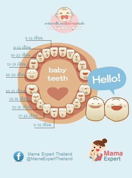 Many of these craftsmen use materials to cover the tooth to preserve and protect them, and at the same time, add their dash of creativity to bedazzle them. This makes for a sentimental yet fashionable piece of jewelry.
4. Shadow box
Your baby’s milk teeth can be a great addition to a shadow box. It can be a great piece of decor in their nursery or room for years to come.
5. Repurposed ring box
If you are looking for the perfect box to keep your baby’s fallen milk teeth, try repurposing a ring box that may be lying around in your house. The folds in the ring box are perfect for holding your little one’s teeth.
1. Can DNA be obtained from an old baby tooth?
Yes. An old baby tooth can be used to obtain DNA. Forensic science now uses bones and teeth to extract DNA from degraded or fragmented human remains for identification purposes. Teeth are preferred due to their location in the jawbone, which provides additional protection to DNA (7).
2. How long do stem cells last in baby teeth?
When dental stem cells extracted from baby teeth are cultured and stored correctly, the cells may remain viable for one to four weeks (8) (9).
Storing a baby’s fallen milk teeth may have emotional or medical significance. For example, parents may save baby teeth as a memory of their childhood, to make souvenirs as gifts to their grown-up children, or for stem-cell retrieval. If your purpose is stem-cell banking, store it in milk till collection or use a preservation kit. Other than that, the tooth needs to be cleaned and disinfected before storage. Later, they may be included in your baby journal or embedded into pendants, lockets, or other jewelry pieces.
Key Pointers
- Some unique reasons to save baby teeth are the much-awaited tooth fairy visit, source of stem cells, and a keepsake for parents.
- You can clean and disinfect them before storing them in tooth preservation kits.
- Some ideas to preserve your baby’s first tooth are a keepsake box, enveloped in a baby book, or you can also make tooth jewelry.


References:
MomJunction’s articles are written after analyzing the research works of expert authors and institutions. Our references consist of resources established by authorities in their respective fields. You can learn more about the authenticity of the information we present in our editorial policy.
1. Irina Kerkis and Arnold I. Caplan; Stem Cells in Dental Pulp of Deciduous Teeth; Tissue Engineering Part B Reviews (2011).
2. P. M. Sunil, et al.; Harvesting dental stem cells – Overview; Journal of Pharmacy & BioAllied Sciences (2015).
3. Benjamin D.Zeitlin; Banking on teeth – Stem cells and the dental office; Biomedical Journal (2020).
4. What Are Tooth Preservation Kits?; Connecticut Children’s
5. Knocked Out Teeth; American Association of Endodontists
6. Should You Bank Your Kid’s Teeth for Stem Cells?; leaps.org
7. Denice Higgins and Jeremy J Austin; Teeth as a source of DNA for forensic identification of human remains: a review; Science & justice: journal of the Forensic Science Society (2013)
8. Caleb Daniloff; Broke a Tooth? Grow It Back; Boston University
9. Mariano S. Pedano et al.; Survival of human dental pulp cells after 4-week culture in human tooth model
The following two tabs change content below.
- Reviewer
- Author
Dr. Meenakshi is a dentist and a passionate writer with over eight years of experience in dentistry and four years in writing. She started her career as a dentist with a dental chain in Mumbai and soon rose to lead the clinic as a Head Dentist. She then switched to working for two start-ups in healthcare, before beginning her own… more
Kay Lakka is the founder of Londontherapy, a busy psychological practise in the center of London. She holds a BSc (hons) in psychology and MSc in the psychodynamics of human development and has numerous post graduate diplomas including advanced psychotherapy, guidance through dreams and psychosexual relationship counselling. Also a doula and hypnobirthing teacher, Kay is a registered member of UKCP,.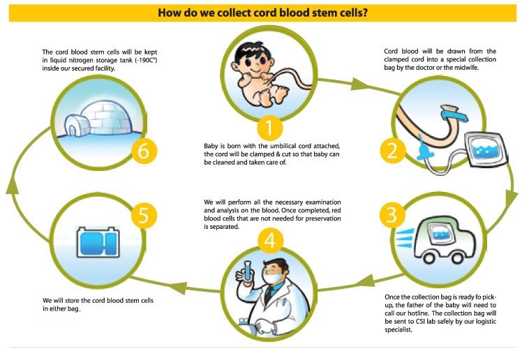 .. more
-
11 Reasons For Your Baby Sticking..
-
11 Reasons For Your Baby Sticking..
-
When Can A Baby Sit In Bumbo Seat? Age,..
-
When Can A Baby Sit In Bumbo Seat? Age,..
-
Hemorrhoids in Babies: Causes, Symptoms..
-
Hemorrhoids in Babies: Causes, Symptoms..
-
Baby’s Head Hot, But No Fever: Causes..
-
Baby’s Head Hot, But No Fever: Causes..
-
Hernia After C-Section: Symptoms,..
-
Hernia After C-Section: Symptoms,..
-
Ovary Pain During Pregnancy: Causes,..
-
Ovary Pain During Pregnancy: Causes,..
-
Bloating During ovulation: Causes And..
-
Bloating During ovulation: Causes And..
-
Leukocytes In Urine During Pregnancy:..
-
Leukocytes In Urine During Pregnancy:..
-
Is High WBC (White Blood Cells) Count..
-
Is High WBC (White Blood Cells) Count..
What Is Stem Cell Teeth Banking
Losing your milk teeth is a natural part of growing up. On average, children in the UK lose 12 milk teeth between the ages of 5 and 10. These milk teeth are gradually replaced by adult teeth, which they will keep for the rest of their lives.
Losing milk teeth is a non-invasive process that children even look forward to as a sign of growing up. Many adults look back to their first visit from the tooth fairy with fond memories, but now the loss of milk teeth has become far more exciting than the expectation of a 50p coin under the pillow.
Stem cell therapy is taking the medical world by storm. The ability to rebuild parts of the human body in a lab in order to replace tissue, tendons, bones and even organs with organic matter that is a perfect match to the individual patient is a real medical breakthrough.
What is tooth stem cell banking?
Tooth stem cell banking. You may have heard of it from your health care provider or even another parent. However, you aren’t sure about what the term actually means, or why it’s important to bank your child’s baby teeth, or even how and where to do it.
Storing stem cells derived from teeth for future medical use is a fairly new practice, although banking cord blood (also a rich source of stem cells) has been around for longer, not to mention the usage of stem cells in bone marrow transplants.
Still not quite sure what any of the terms mean? This guide aims to explain what they are, and to take you through medical tooth storage, from start to finish.
What is a stem cell?
Stem cells are the body’s building blocks. They are able to multiply themselves to create new cells and change into whichever type of cell the body needs. A single cell from a child’s milk tooth is capable of adapting itself to become bone, muscle, tendons, skin, nerves and cartilage.
Cells – more specifically stem cells – are essentially the building blocks of our bodies.
The cells in our body are able to replicate themselves (a skin cell is able to form other skin cells, blood cells will divide into other blood cells, and so on). However, stem cells are able to reproduce themselves not only into other stem cells but also cells capable of growing into any other cell in the body under certain conditions – almost like blank slates. This has led to a lot of promising research into regenerative medicine. It means advances into treating illnesses such as diabetes, Crohn’s disease, Parkinson’s and spinal cord injuries, to name just a few.
The treatment process when using stem cells is similar to blood marrow transplant. A stem cell transplant allows your body to grow healthy cells stemming from the original to repair or replace damaged ones.
Typically, stem cells are harvested from umbilical cord blood from newborns and from milk teeth in children, although they can be found in adults as well. They are located in a few places across the human body, but exist in greater numbers in dental pulp, thereby ensuring better viability when the time comes to retrieve them.
Harvesting stem cells
How do you harvest stem cells from teeth?
Stem cells, in general, are not easy to come by. Until now, the most common way of harvesting stem cells to use in stem cell therapy has been by removing part of a patient’s bone marrow and isolating the stem cells in a lab. In this procedure, doctors usually use a long needle to withdraw bone marrow directly from the pelvis and backbone. The patient must have a general anaesthetic before undergoing this procedure, otherwise, it would be extremely painful. The patient would then usually need around a week to recover while their body creates enough bone marrow to replace the quantity taken.
Many people are aware that stem cells are stored in the bone marrow in the body, but few realise that there are also stem cells present in milk teeth. Harvesting stem cells from teeth is much less invasive for the patient than extracting bone marrow. Stem cells can be found in dental pulp, which naturally occurs inside milk teeth. This dental pulp is taken to the lab and stem cells are isolated in the same way that they would be from a bone marrow sample, but, seeing as losing milk teeth is a natural part of life, this is much less traumatic than the bone marrow alternative.
The lack of an invasive medical procedure required to procure a dental stem cell is great for a number of reasons. Firstly, there is no risk of harming your child or affecting their health in any way, making it a safe, rather than daunting, procedure. Secondly, far fewer medical staff and pieces of medical equipment are required, making the process much cheaper than alternative ways of harvesting stem cells.
Why should you save your child’s baby teeth for stem cells?
Life is unpredictable. Illnesses and injuries happen, not to mention just plain growing old. Stem cell banking is a form of insurance for your child’s future. Imagine a sort of restart point for the cells to regenerate from again in the event of misfortune, or even just simply age. Perhaps we can’t regrow things like entire limbs presently, but there is good evidence that we can, and have regrown cells for other purposes, like knee cartilage repair for when your child grows (much) older, or for blood-based illnesses like HIV/AIDS or certain blood cancers. For now, medical research is still ongoing, and it is strongly believed that in the near future, stem cells will be used in ever more wide-ranging fields, which is a good reason to preserve stem cells while they are young.
Aside from ensuring that your child has healthy cells to fall back on later in life, the same stem cells will also be able to help blood-related family should they fall ill with life-threatening diseases.
Stem cells harvested from children’s teeth are generally of better quality than when they are taken as adults since they are less affected by factors such as ageing, accumulated illness or environmental factors, which can lead to cells of deteriorated quality. As they grow older too, cavities may also affect whether a tooth is available to be used for stem cell banking.
Can baby teeth stem cells be used to heal and repair from illness?
Once the stem cells are harvested and scientists are able to get them to multiply into enough cells to start building tissue, they can begin to be used in stem cell therapy. This medical technique is on the rise as more research is being carried out into exactly how far it could progress the course of medicine.
As it stands, we know that stem cells can be used to create skin grafts to help patients recover from severe burns, rebuild the connections between nerves to help combat brain damage, rebuild cardiac muscles damaged by heart disease, create cells which secrete insulin to cure diabetes, replace cartilage, tissue and bone and potentially treat patients suffering from Parkinson’s, Alzheimer’s and other degenerative motor neurone diseases.
The extraordinary power of stem cells to transform themselves into other cells within the body means that the possibilities will only continue to increase as more research is conducted.
Store a tooth UK
Why store milk teeth?
Storing your child’s milk teeth will give them the best chances should they need stem cell therapy in the future. As this field of medicine grows and develops, the possibilities for the power of stem cells to cure more and more conditions will only increase. The knowledge that your child’s stem cells are already stored will give you peace of mind, knowing that, should your child be injured or develop a serious condition, you’ve done everything you can to give them the best shot at stem cell therapy.
There are no guarantees, but having the stem cell teeth stored and ready to use will give your child the best fighting chance if the doctors think that stem cell therapy is a viable treatment.
Why do we use stem cells in teeth?
Stem cells collected from teeth are just as viable as those sourced from bone marrow. Seeing as everybody loses teeth anyway, it is an excellent way of banking stem cells for your child and your family as a whole and storing them, in case anything should happen in the future.
Baby teeth storage
Is it easy to save baby teeth?
For some families, the decision to save their child’s milk teeth could not be easier. There are a large number of genetic diseases passed on from generation to generation that are starting to respond to stem cell therapy. Parents who know that they carry genes for a particular hereditary disease, such as sickle cell, or whose children already have a diagnosis for such a condition, are saving their children’s milk teeth to give them the best possible future. By ensuring that their child’s stem cells are stored when they are most viable, parents are doing everything they can to increase the chances of successful stem cell treatments in the future.
Tooth banks
What is a tooth bank?
Tooth banks are a type of medical storage unit which takes in teeth from donors all across the country. There are laboratories onsite which process the teeth and extract the stem cells before making sure that they are ready for storage. The tooth bank will then take care of the milk teeth stem cell storage so that the cells are ready to use should you ever need them.
How do you extract stem cells from a tooth?
Teeth are fully inspected to check that they are healthy and in good condition when they arrive at the laboratory before the technician can begin to extract the stem cell from teeth. The tooth is disinfected and the dental pulp is extracted from the main tooth. The dental pulp is the soft tissue that is found inside teeth. It is the part that receives a blood supply from the gums and is also the part of the tooth which stores the stem cells. This soft tissue and the teeth cells inside are treated with what is called a cryoprotectant, to help preserve the cells during freezing. The cells are then kept in medically inspected freezers, where they will stay until they are required.
How long can you store a tooth for?
To date, the longest recorded storage period for stem cells, once they have been extracted, is 22 years. Researchers found that after 22 years, the cells were still as healthy and as ready to use as they were when they were first harvested. Other types of human cells have been successfully stored for 50 years.
What’s the difference between cord blood banking and dental pulp banking
Cord blood and dental pulp hold two different kinds of stem cells. Umbilical cords hold hematopoietic progenitor cells or HPCs. These are used primarily in the treatment of blood diseases – leukaemia and lymphoma, for instance.
Tooth stem cell banking is a newer process, having only been around since the 2000s. The cells found in teeth are known as mesenchymal stem cells or MSCs. One may also find MSCs in bone marrow, but they are inferior in quality compared to the ones found in healthy teeth. They have the potential to be used in treatments for several other types of tissue and organs such as muscle and skin, as opposed to the more limited usage of HPCs in blood diseases.
One notable recent development in the use of MSCs in therapy involves the growth of new skin for burn victims, via the spraying on of stem cells to the affected areas. The results have been encouraging. Another is the use of stem cells to regenerate badly damaged lung tissue, in the wake of respiratory pandemics in 2009.
Why save baby teeth for stem cells?
Stem cells from baby teeth are particularly prized because there is evidence to suggest that stem cells from younger donors have more of a success rate when it comes to medical procedures. The younger the milk tooth, the more potent the stem cells inside it, so, for the most viable stem cells, baby teeth are required.
There is also the added bonus that donating dental stem cells requires no special surgery or any particular effort. You simply let nature take its course and wait for your child to lose a milk tooth, rather than having to subject them to an invasive medical procedure. It is then extremely easy to order one of our kits, follow the instructions to package and send the tooth, and we’ll take care of everything else.
How to store milk teeth
It is an easy process to store dental pulp with Stem Protect. You can make an appointment even before your child’s milk teeth are beginning to come out.
First, fill out a contact form that you can find on our website, or give us a ring at 0115 967 7707. We will send you a storage agreement for you to sign, and we will also require the initial payment at this point. After that, a collection kit will be mailed to your home, and one of our staff will let you know your phlebotomist’s details by email as well.
When your child’s tooth falls out, follow the instructions in the collection kit closely. Once that is done, please arrange for a courier to come to pick it up soon, and make an appointment for our phlebotomist to come by for a blood sample. We have collection services 365 days a year, so rest assured that you will be able to send the tooth to us any day, even if it is a weekend or holiday.
That’s it! Now your child’s milk teeth will be processed and stored safely for the duration of the agreement that you sign for. They will be kept in several samples in a highly secure bank to ensure zero worries on your end, and the ability to use them multiple times if needed.
At our lab, all dental pulp will be carefully and cryogenically preserved at temperatures of -150 degrees Celsius. So far there is no reason to believe that there is an upper limit to how long the tooth stem cells can be stored and still be viable even after thawing; this means that as your child grows up and ages, their cells will still be available in the event of any treatment or therapy they require.
How much are tooth banking costs?
Choosing to store your teeth or your children’s teeth is an elective procedure and, as such, it is not covered by medical or health insurance. Prices for storing your children’s teeth will depend on the package you choose. There are different options according to how many teeth you would like us to process and how long you would like us to store them for. If you give us a call or contact us through our website, we would be more than happy to discuss pricing options with you.
The actual cost will vary based on the length of time that the teeth are stored for. Generally, prices are reasonable and affordable. Right now Stem Protect offers an annual plan as well as a 25-year plan. You can opt to pay a one-time fee or in instalments. Please contact our friendly customer care team to learn more.
Extra services
We understand that the storage of your stem cells is important to you, and that is why we have a business continuity guarantee. No matter what happens, rest assured that your cells will be well protected.
For the new mother and the baby, we provide both prenatal and newborn screenings, along with diagnostic services for life-changing conditions. These can include things like Patau’s Syndrome and coeliac disease. Alternatively, we are also able to collect and preserve umbilical cord blood for babies.
Adult tooth stem cells banking
Can you store adult teeth stem cells?
If you’re an adult and wish to store your stem cells with us, we provide banking services via dental pulp and adipose tissue.
To date, Stem Protect has processed and stored over 125,000 samples for upwards of 75,000 families. We are an accredited and reliable stem cell storage company in the UK, and we process more samples than any other bank at the moment. Call us today to make an appointment, or for a free, non-obligatory consultation.
What to Do With Your Child’s Baby Teeth
It is surprising to see a baby become a toddler, then a kid, and see them looking funnily toothless after losing their baby teeth. In fact, an emotional moment for parents is saying goodbye to baby teeth, as it marks kids getting into the last part of childhood.
Also, sometimes seeing a kid losing their teeth takes parents by surprise. As a result, parents constantly ask us questions about what to do with baby teeth? Thinking of this, we have created a list of baby tooth-related subtopics based on common parents’ concerns.
With this brief introduction, we then deal with folklore myths scaling to transcendental topics such as the importance of keeping baby teeth and its relation to stem cell research and potential future clinical needs.
The Traditions to Dispose of Teeth.
Historical depictions relatable to baby teeth traditions that occur around the world are pretty entertaining to read. For instance, a common custom that has survived to these days is the tooth fairy tale.
The history of the tooth fairy has passed generations. Parents tell their kids the story that a Tooth Fairy will appear at night while their kid is sleeping and take the tooth that has fallen, leaving money or other treats in exchange.
The magic occurs only if a kid leaves a baby tooth under the pillow. But intendedly, parents use this magic to ease kids’ fear of losing a tooth.
Europe
People have replicated this tale inadvertently for centuries. Early narratives describe a Norse tradition characterized by superstitions where a single possession like a tooth control forces of nature or uncommon events.
People thought kids’ teeth bring good luck, so Viking warriors made baby teeth necklaces or buried a tooth, believing that it would help the kid resist the struggles of an afterlife. Later, the parents left a small fee for the teeth’s favors.
During medieval times in Europe, parents tossed baby teeth into the fire. They believed that by doing so, they would free their kids from the malign manipulative forces of witches.
Now, back to the Fairy Tale Tradition, a priest wrote an enchanting story in Spain in 1894, when King Alfonso XIII, an eight-year-old child, saw his first tooth falling off. So Queen Maria Cristina appointed father Luis Coloma Roldán to write a story to calm her scared kid.
The tale tells a heartwarming story about King Bubi transformed into a mouse. Little Ratón Pérez was King Bubi’s companion and guide. Little Ratón Pérez revealed to Bubi the daily struggles the crown subjects faced.
The story’s primary purpose was to teach King Alfonso XIII values like kindness and bravery. Later on, the story was adapted and publicized, appearing in Wisconsin in 1950. Little Ratón Perez’s popularity was the germ for adaptations in Japan, Russia, and China.
Other baby teeth traditions relate to burying a kid’s first baby tooth in the place where parents wish their kid will develop its associated attributes. In Turkey, for instance, parents might choose a soccer field, expecting the kid to be a soccer superstar.
Asia
In Asia, people throw baby teeth away, believing this will boost the growth of healthy permanent teeth. Also, children throw their mandible teeth to the roof of the house, expecting their new teeth to grow upward, and bury the maxillary teeth as profound as possible, wishing their teeth to grow downwards.
In short, baby teeth are the source of traditions, seen as a material source of power against undesired events, and have also helped to create literary work. However, few could deny that a baby tooth has sentimental value for parents.
For this reason, we now want to share with parents some suggestions about what they can do to keep baby teeth and how they might serve a fruitful purpose, caring for your child’s health.
How to Preserve Baby Teeth?
We have an assortment of plans you can revise and choose from if you decide to preserve your kid’s baby teeth. Notwithstanding, there are some facts you might want to revise with us about adequately storing and keeping teeth.
Here is a three-step process for keeping a baby tooth for a long time:
Clean
You just have to rinse-soap-rinse the tooth in abundant water and soap.
Disinfect
Use rubbing alcohol on the tooth’s surface.
Dry
Use a clean towel or air dry the tooth.
Now we are ready to talk about what you can do with your baby lost teeth.
What to Do With Saved Baby Teeth
It’s very interesting, but this Dentavox Infographic shows some of the preferences on what to do with baby teeth. Once children’s teeth are taken away by the Tooth Fairy (and totally not their parents), the question is about what to do with them.
Even if nearly 3 in every 4 adults don’t have their primary teeth stored anywhere, over half of those surveyed stated they would like to save their children’s teeth.
Maybe it’s due to a feeling of regret; after all, the number of people saying they regretted not saving their teeth was nearly in the same proportion as those who claim they would like to save their kids.
That has to say something about our upbringing, right? We’ll leave that to the investigators in the appropriate field.
Some of the most popular reasons why adults decided to save their children’s baby teeth included:
- Following family traditions (even if it seems weird to you, some of those traditions are very nice)
- Trying to make the children happy (even some kids ask to save their primary teeth)
- They saw it as the most practical solution (we are not exactly sure as to what was the original problem, but we’ll take the help we get.)
From the minority stating they would throw away the teeth, some also claimed this meant following family tradition, following some type of ritual. A few individuals also mentioned they chose to bury them as the preferred disposal method.
Keepsake Box
Our kind suggestion is to have a specially dedicated box to preserve your kid’s baby teeth. You can find a keepsake box in the form of a hearth that resembles how much you appreciate the tooth you want to preserve.
Another option is to provide a new purpose to an existing item you love, like a jewelry box. Though, it might be too small to keep more than one dental piece, so we have some other options for you.
You can surf the web and find some keepsake boxes intended to put every single baby tooth in a purposely designed spot. For instance, Etsy has an assortment of options you can check by scrolling down to find the one you like the most.
Baby Book
Many parents opt to have a baby book to save pictures and the most valuable things that marked their baby’s attainments. In addition, a baby book can bring enjoyable memories to parents’ minds by keeping their baby teeth in the baby’s book. An easy way is to attach an envelope with the tooth and assign it a page with the date it fell.
Tooth Jewelry
This does exist, and believe us, it is not disturbing. In fact, a baby tooth falling is an emotional moment parents wish to preserve. You can also have custom-made jewelry design charms with the tooth as the main piece.
There is a lot of space for imagination. You can try getting an earring or a necklace, and why not? You can also have a ring designed for your preference.
Science Projects
A wise choice is to save your kids’ baby teeth for their use in an elementary school science project. For instance, your child can prove the unwanted effects of corroding acids present in sodas over teeth enamel. By the way, now that we have touched on science as a topic, why don’t we revise what we consider the most relevant option parents can opt to do with baby teeth.
Save Baby Teeth for Stem Cells
To start defining the importance of saving baby teeth for stem cells; we found it relevant to describe a systemic disease. When we refer to the word systemic, it affects the body as a whole instead of a single organ or part—for instance, having high blood pressure.
With this said, Stem Cell treatment might be crucial in solving specific systemic disorders or diseases that might present in the future. However, we can’t ignore the fact this type of treatment is expensive because it is fairly new in the healthcare world.
Harvesting stem cells from adults is painful because it requires doing so from bone marrow, but recent studies have proven that the scientific community can also harvest stem cells from teeth. Another study highlights the usefulness of teeth-harvested stem cells.
So, before thinking of disposing of your kid’s baby teeth, please think twice. Medical technology advances tremendously rapidly, and saving and preserving baby teeth can make a massive difference to your loved one.
Finally, preserving teeth for this purpose requires special treatment, so we encourage you to entrust your kids’ baby teeth to qualified experts so that they can treat and harvest stem cells from them. Then, call us so we can help you send the teeth immediately after they fall out.
What Is the Timeline for Baby Teeth to Fall?
By the age of three, children develop their primary teeth. However, some of these teeth stay for a long time until the teenage years. Also, although this set of teeth tends to fall, its care influences the development of permanent teeth that are about to erupt.
Consequently, we encourage you to educate your little kid about cavities, decay, and gum disease and how to prevent them with excellent child oral care. Fortunately, children grasp and process formative messages by the age of three.
Back to the point, the timeline for teeth to fall and erupt ranges between 6 and 21 years old if considering the third molars (wisdom teeth). We include a timelapse shortlist of events related to baby teeth falling and permanent teeth sprouting.
Age 6:
First in, first out. The first teeth that appeared fall also first by this age. You might also expect to see the first molars erupting in the back of the gums.
Age 8:
Front upper and lower teeth (central and lateral incisors) fall, and their replacement is their permanent counterparts.
Ages 9 to 10:
You might not see any disruptive changes during this time.
Ages 11 to 13:
By this age, the rest of the teeth should have fallen, including the canines, also known as cuspids, and the first and second molars. Their replacements come along immediately after.
Ages 14 to 17:
You might not see any disruptive changes during this time.
Ages 17 to 21:
Patients might not see or even feel it, but the last set of molars (third molars), also known as wisdom teeth, might erupt or develop impacted, meaning there is no space for them to sprout. Impacted wisdom teeth might stay below the gums or bone or partially erupt in an angled position. There is also a possibility that wisdom teeth never appear or only some of them show on.
Do Falling Teeth Require Special Care When Loose?
We all had wiggled and played with loose teeth when we were a child. Actually, we can’t deny it’s fun to make videos, take pictures, or joke around falling teeth. However, tooth falling is a natural, painless process, so applying unnecessary force to a tooth that is not quite ready to fall might damage tooth roots and lead to an infection.
What if My Child Is Late Losing Her Teeth?
Discard any major concerns about late falling teeth. The timelapse presented above is an approximation to a tooth’s baby falling. However, as in any other physiological process, no person’s response is similar.
The time the first tooth sprouted might influence the teeth falling process. Babies having their first teeth soon will eventually have their teeth falling soon as well. The same condition applies to late baby teeth receivers.
My Child’s Baby Tooth Has Fallen Off, What Should I Do?
Once you have your kid’s first baby tooth in your hand, you start wondering what to do with it. Should I keep it? Why is it important to keep it? Or maybe by following a tradition, you might opt to discard it.
Incredibly, there are plenty of choices, and critical decisions might come from a single tiny denture piece. But, we know and understand that kids don’t come with a manual, so as parents, we must wisely evaluate the most appropriate option.
Consequently, far from any suggestion about what to do with your kid’s baby teeth, we can provide you with some alternatives you can choose from to do with these teeth. We include all sorts of possibilities.
However, before entering into detail, we sensitively suggest parents keep their kids’ baby teeth. This is because medicine advances overwhelmingly rapidly, and baby teeth might be a fundamental resource for medical treatment.
Why Do Some Adults Keep Baby Teeth?
We have a thought-provoking fact. This infographic depicts some of the most typical representative actions done with baby teeth. Even though most parents don’t keep their baby teeth stored, approximately half would like to save their kids’ baby teeth.
Research findings unveil the reason why parents would opt to save their children’s baby teeth, and they are:
A Family Tradition
We can leave that to a cultural or solely a bonding custom, but we find them plausible and, of course, tender.
It Is Entertaining for a Kid
Why not? This is an outstanding event for a kid.
It Is Practical
Parents might have their thoughts about what could be the concern, but we will try to investigate further in this article.
Reasons to throw away and dispose of the teeth include traditions and even rituals. Additionally, some parents just find burying teeth practical. Whatever the choice parents make with baby teeth, the truth is, customs, stories, or narratives sometimes guide parents’ actions regarding their child’s teeth. Whether they are good or bad, we will revise some of them.
Baby Teeth Myths
We love the internet. We can get all sorts of interesting and educating information there. But unfortunately, you can also be misguided with poor and sometimes exaggerated content. Also, myths transcend from mouth to mouth, forming beliefs.
Myths are just widely held but false beliefs or ideas and should be understood this way. However, sometimes myths transcend objectivity and are taken as certainties. Myths might confuse parents leading them to neglect kids’ dental care.
Therefore, we want to thwart some of the most common beliefs that cause confusion while entertaining parents with some weird misconceptions about baby teeth and telling them what not to do with them.
Myth N° 1: Baby Teeth Aren’t Important
When we talk about baby teeth, we refer to the entire development of baby teeth from the moment they sprout up to the point at which they fall. So, neglecting their importance is disregarding the complexity of future mature teeth formation and their role in adults’ life.
To clear things, people tend to believe baby teeth will just fall, so why would they care about them while they are functional. First, they hold the space for the entire dental structure to come, help in the progression of speech, and allow kids to mature eating and masticatory habits that will be fundamental as they grow.
Most importantly, baby teeth serve as natural guides for newly coming permanent teeth preserving the natural separation so they follow a pattern. Unhealthy or neglected baby teeth might drift into permanent sprouting teeth.
Thinking ahead, a lost baby tooth due to a cavity or an accident might derail the upcoming tooth’s development, leading to orthodontic problems like crowding affecting other teeth, making them hard to clean.
Untreated or neglected baby teeth might derive from costly treatments to realign them. For this reason, we recommend you attend with your kid to pediatric dental care for checkups to promote proper teeth development at early stages and ages.
Finally, a missing tooth negatively impacts a child’s masticatory motion. As a result, poor mastication deprives kids of fully absorbing food nutrients ending in developmental and health-related problems.
Myth N° 2: There Is No Need to Fight Cavities in Baby Teeth
Myths sometimes share a source, and this is one case of it. The last myth relates to the misconception that there is no need to fight cavities in baby teeth. Once again, preventive measures to keep teeth healthy are a must.
Studies collected from the Centers for Disease Control and Prevention (CDC) revealed that almost one-half of kids in the United States with ages between 2 and 9 had suffered from at least one form of tooth decay.
Tooth decay can develop into dental caries. Bacteria penetrate the enamel shield of teeth, making them vulnerable. Untreated caries permit bacteria to advance, causing pain and producing infections that can be spread through the blood vessels, affecting other body organs.
Cavities are a genetic thing. This is not a myth but an overused argument. Some people tend to think that cavities are something they can’t fight against. Now, this is a myth.
Despite the minimal influence genetics have on the development of cavities, they are absolutely controllable with adequate oral hygiene habits. So, we encourage parents to educate and guide kids on brushing and flossing after meals.
Also, parents have a mission to take theguir kids to checkups and ask a pediatric dentist in Katy about treatments that include sealants and fluoride applications to reduce the propensity to develop dental caries in kids.
Bonus Myth: Placing Aspirin on a Toothache Will Alleviate the Pain
It might sound odd, but this myth does exist. We don’t know where this recursive fervor for storytelling comes from, but it is our job to null this erroneous statement. Put simply, aspirin does not work this way.
Aspirin blocks certain chemicals that transmit the sensation of pain. To do so, aspirin must flow through the bloodstream, and the intestine absorbs it. The mere fact of placing an aspirin on a kid’s tooth will bring no relief whatsoever.
Instead, if your kid complains of a toothache, we are called to recur to a pediatric dentist immediately as parents.
Myth N° 3: There’s No Need to Brush Baby Teeth
We are happy to talk and analyze this statement. So we include a definite consideration, please note: “parents should brush their kids’ teeth right after their first teeth sprouts.” The previously thwarted myths lead us to vindicate brushing and flossing.
Going a little further, we encourage parents to start caring about their kid’s oral hygiene before their first tooth sprouts. For instance, you can use a damp rag and rub it against your kids’ gums to eliminate any trace of food that can serve bacteria as a breeding source.
Also, parents refrain from brushing and flossing their kids’ teeth to avoid alarming them when they see their gums bleeding. In such circumstances, we encourage parents to use a soft-bristled toothbrush and continuously reinforce teeth brushing’s importance. If your kid’s gums keep bleeding, consult a pediatric dentist for an evaluation.
Myth N° 4: Kids Don’t Need to See a Dentist Until They Are Older
First-time parents subdue to the thought that the first pediatric dentist visit should occur once they find a dental problem with their kid. But, as the American Academy of Pediatric Dentistry (AAPD) suggested, parents are encouraged to take their kids to their first dental consultation at year one or immediately after their first tooth sprouts.
Parallelly, by showing your kids that the pediatric dentist’s office is a fun place to be, you are helping to develop a calm instead of a tense sense about dentists, forming a trusting relationship that nulls the reluctance to go to a dentist.
Myth N° 5: Adults Cannot Have Baby Teeth
Yes, adults can have baby teeth. In fact, this is a common diagnostic that is also known as retained teeth. The most prevalent case of retained teeth is when there is no permanent replacement tooth growing.
Specifically, a study shows that retained second molars are less likely to produce a future dental problem by age twenty. On the other hand, this is not the case for incisors and first molars, as they might require particular intervention.
Concurrently, adult baby teeth should not be left unattended as a neighbor tooth can’t erupt appropriately because baby teeth remain in a fixed position. Also, there might be cases of a misaligned baby tooth when closing the mouth, and finally, a retained tooth might cause a space between teeth.
When Should You Brush Your Child’s Teeth?
We previously commented about the perfect timing to start brushing your kids’ teeth. Furthermore, there is a misconception that brushing right after a meal might damage teeth. The truth is, we should revise this all together in detail.
For instance, if your kid has delighted themselves with an orange, this fruit contains citric acid that can wear enamel. However, saliva serves to wash unwanted residues in the mouth. So, you can wait an hour and then assist your kid in brushing her teeth for at least two minutes.
FAQ
What to do with baby teeth?
Some people discard baby teeth, others hold on to them. For those of you who are thinking of keeping your child’s baby teeth after they have fallen off, you can clean them well and put them in keepsake boxes, turn them into jewelry pieces, or save them for your child’s future science projects.
What to do with tooth fairy teeth?
If you decided to keep your child’s baby teeth after they fell off, you can do a lot with them. Try saving them for a future science project to show how different drinks can affect them in the long term.
What to do with baby teeth after they fall out?
Many parents like saving baby teeth as a reminder of those first years with their child. Some parents like preserving them in a keepsake box, and even others like integrating them into pieces of jewelry like collars.
How to preserve baby teeth?
The first step in keeping your child’s baby teeth is cleaning them thoroughly. You can start by cleaning them with soap and water, but also remember to swab them with alcohol to completely disinfect them. Dry them well and keep them away.
How long can you keep baby teeth?
Baby teeth won’t deteriorate much if you keep them away. Of course, there are other means to preserve teeth depending on the use you have for them. Some parents may want to keep them for stem cells in case of the need for some medical treatments, but this requires other specialized resources.
Banking on Baby Teeth: Dental Stem Cells and Regenerative Therapies
A month ago, my 6-year-old wiggled her first tooth out, and the tooth fairy dutifully left a glitter-strewn $1 bill and a nice note. In response to my Facebook post announcing this major milestone, my mom pointed out an article about “banking” baby teeth because — get this — the living dental pulp inside baby teeth contains stem cells.
“Stem cells” might ring a bell for women who delivered babies in a hospital or birth center, because most of us were asked if we wanted to store or donate the stem-cell-rich umbilical cord blood. Stem cells are the body’s biological wild cards, with the potential to be transformed into a variety of other cells and used in medical therapies to replace damaged or malfunctioning cells. Think of it as a way to treat an ailment at a cellular level specific to the individual, rather than just treating symptoms.
For that reason, many parents decide to “bank” their baby’s umbilical cord upon birth.
“Up to 40 percent of qualifying mothers with normal term pregnancies opt to donate cord blood to the public bank, and private donation is even more frequent,” says Dr. Rebecca Haley, medical director of Bloodworks Northwest. Last year alone, 250 units of publicly banked cord blood were sent for transplant through the Cord Blood Coordinating Center and used in treatments for leukemia, lymphoma, rare cancers and metabolic conditions.
But back to the baby teeth. Growing up around my dad’s dental office, I saw and learned some fascinating things, but stem cells inside teeth? It blew my mind to think that my child’s baby tooth could hold the key to a life-saving treatment in her adulthood.
Where the tooth fairy banks
To collect and store dental stem cells, a dentist must extract the baby tooth when it starts to get wiggly and then prep it with materials from a special kit provided by the chosen dental stem cell bank. Currently there are five such banks located in the United States. Once the doctor preps the tooth, it’s sent overnight to the chosen bank, where, upon confirming the cells’ viability, they’re cryogenically preserved (i.e., frozen) until needed.
It blew my mind to think that my child’s baby tooth could hold the key to a life-saving treatment in her adulthood.
Currently, this relatively new service is only available privately, which means you have to pay a one-time processing fee that varies from $500 to $1,700, plus an annual storage fee of about $100 to $200. To differentiate themselves, some labs tout higher lab certification standards, options to duplicate cells to enlarge the specimen sample or provide an environmentally friendly processing kit to the dentist. Most labs also affiliate with larger ones, in case the business should change hands or something happens at the storage site.
Cord vs. teeth
But aren’t all stem cells the same? Isn’t donating your baby’s cord, if you choose to do so, enough? Not exactly. There are important differences between dental (mesenchymal) and umbilical cord (hematopoietic) stem cells. Dental stem cells can become, among other options, bone or muscle cells to treat issues associated with those areas of the body, much like doctors already use umbilical cord stem cells in blood-based therapies to regenerate blood and bone marrow for cancer patients.
Also with dental stem cells, you have at least 24 chances (that’s the number of baby teeth plus wisdom teeth) to gather them over the years your children lose their teeth. These cells can also be duplicated on a massive scale, so even a small viable sample can theoretically yield a large bounty. Conversely, with umbilical cord stem cells, you get just one chance to gather them — at birth. The number of cord stem cells you get is all you get, as there is not yet a method for duplicating them. There are, however, public banks where people can donate or receive umbilical cord stem cells.
“Biological insurance”
So why is it that you haven’t heard of dental stem cell banking? It’s still a work in progress, with many treatments and therapies under development. The U.S. Food and Drug Administration has yet to approve the widespread use and application of dental stem cell therapies, with only animal studies and limited clinical human trials conducted thus far. Advocates are hopeful that the successes with umbilical cord stem cell therapies will hasten the approval process for dental stem cell therapies within the next decade.
Think of banking dental stem cells as “biological insurance,” says Arthur E. Greco, CEO of StemSave, a dental stem cell bank in New York City. He and other supporters of dental stem cells believe regenerative therapies are poised to revolutionize medicine.
“Young people today are projected to have life spans of 100-plus years,” says Greco. “Regenerative therapies will play a central role in assuring that those longer life spans will be healthy as stem cell treatments are utilized to combat the normal degradation that occurs as we age.”
While this may sound like science fiction, medicine is moving toward, customizing therapies and medications down to the cellular level. There is still much work and research needed, but by the time our kids hit middle age, this type of treatment could be a distinct reality.
“This area of study is moving quickly, and significant clinical applications may be available in the future,” says American Academy of Pediatric Dentistry national spokesperson Dr. Amr M. Moursi. “Parents should discuss the risks and benefits of dental stem cell banking with their pediatric dentist in order to make a well-informed decision.”
While it’s not a decision to take lightly, Seattle pediatric dentist Dr. Purva Merchant embraces dental stem cell collection. “Stem cells are becoming more and more invaluable in retaining genetic information that is specific to that particular individual,” she says. “This will help in customizing medication for certain genetic conditions.”
If you’re interested in dental stem cell banking, read up on all of the options and find the one that best fits your needs and budget for the long haul. After all, you’re setting up a potential option for your children’s medical well-being that will follow them into adulthood. While some parents may be ready to jump on the dental stem cell bandwagon now, others might want to wait and keep tabs on future medical research partnerships and FDA trials. Either way, I bet you’ll never look at a loose baby tooth the same way again. I know I won’t.
The tradition of keeping milk teeth was supported by geneticists
The science
11531
Share
Milk teeth contain dozens of valuable stem cells suitable for growing new organs and treating various diseases. This conclusion was made by Dr. Suntao Shi from the American National Dental Institute (National Dental Institute).
Photo: Gennady Cherkasov
A team of experts led by Shi found that adult teeth contain only one type of stem cells, while children’s milk teeth (up to 8-10 years old) consist of completely different stem cells. They are located inside the tooth, in the pulp. With the help of these cells, in the future it is possible to restore tooth tissue or grow pancreatic cells that produce insulin. The main thing is to send lost milk teeth for storage in time.
According to the doctor, a child’s milk teeth should be frozen within the next 48 hours after they fall out in special storages – then there will be a guarantee that the cells in them will be preserved.
Comment by the Head of the Laboratory of Neurogenetics and Developmental Genetics of the Institute of Gene Biology of the Russian Academy of Sciences, Professor of the Russian Academy of Sciences Galina PAVLOVA:
Yes, there are indeed stem cells in milk teeth, or rather stem and progenitor cells, but you still cannot save them yourself. In our country, there are banks of cord blood and placenta, but there is no storage of milk teeth. Of course, extracting stem cells from milk teeth is the most painless procedure compared to other methods of extracting stem cells, but, firstly, this procedure is much more complicated than extracting them from umbilical cord blood, and secondly, the stem cells themselves in milk teeth are not so much. Cultivation is required to obtain enough of them, and long-term cultivation associated with uncontrolled division entails a genetic change in cells. That is, before starting cultivation, it would be necessary to develop a technology for monitoring possible changes. In general, stem cells from teeth have great potential for further use: they can be used to grow new teeth (there are already such technologies in the West), blood vessels, and nervous tissue. Maybe now, in connection with the federal law “On Biomedical Cellular Products” adopted in the summer, we will have more opportunities for studying stem cells, and there will also be jars for storing milk teeth.
Subscribe
The authors:
-
Maria Bykova
Children
What else to read
What to read:More materials
In the regions
-
Putin announced partial mobilization in Russia: who will be affected
42129
Ryazan
Anastasia Batishcheva
-
Residents of Ulan-Ude become prostitutes to pay off debts and help relatives
26098
Ulan-Ude
Roxana Rodionova
-
“There is no girl – there is nothing to lose”: what happens in the military registration and enlistment office of Barnaul on the third day of mobilization
15416
Barnaul
Anastasia Chebakova
-
The Magnitogorsk Drama Theater told about the director Sergei Puskepalis, who died in an accident
12434
Chelyabinsk
Albina Khokhlova
-
Kostroma problems: mushrooms disappeared in our forests
10727
Kostroma
-
“We need to tune in”: a stylist in Ulan-Ude predicted the return of the zero years fashion
A photo
7760
Ulan-Ude
Seseg Zhigzhitova
In the regions:More materials
MILK TEETH WILL HELP ADULTS | Science and Life
Medical and scientific interest in stem cells is based on the desire of mankind to find a source of new, healthy tissues for the treatment and restoration of damaged organs, including those whose loss seemed irreparable before.
Science and life // Illustrations
Obtaining embryonic stem cells and ways of their subsequent differentiation.
The dental pulp, the pulp, is made up of connective tissue and provides nourishment and growth to the teeth. The pulp is in a “case” of dentin, which is a type of bone tissue. Enamel and cementum cover the dentin, thereby protecting the dental tissue from wear.
Stem cells isolated from the bone marrow during transplantation provide restoration of damaged bone tissue of the face and jaw, and cells obtained from the dental pulp restore damaged dentin.
‹
›
View full size
STEM CELL METAMORPHOSIS
The precursors of all the cells that make up an organism are called stem cells. For the first time, the existence of stem cells was suggested and proved at the beginning of the 20th century by the professor of the Imperial Military Medical Academy A. A. Maksimov.
Absolutely all cells of organs and tissues have a single “progenitor” – the so-called multipotent stem cell. In the process of division, two daughter cells are formed from one stem cell, of which one is identical to the mother cell, and the other can produce a variety of cell progeny that is different from the precursor cell. All stem cells have the ability to reproduce their own kind and can turn into several types of cells. This property is called multipotency. From generation to generation, stem cells lose their multipotent properties, acquiring the ability to turn exclusively into cells of one or several tissue types, and eventually become the “building blocks” of tissues of a certain type.
In humans, stem cells have been found in embryonic tissue, some fetal tissues, the umbilical cord, placenta, and adult tissues and organs, such as bone marrow. These cells play an important role in the repair of damaged tissues. For example, as shown by recent animal experiments, only 10 stem cells are able to completely restore hematopoiesis within a few weeks after transplantation of cell culture into the bone marrow.
There are four main types of stem cells: embryonic, fetal, somatic and mesenchymal.
Embryonic stem cells are found at the earliest stage of embryonic development. A fertilized egg (zygote) begins to divide 30 hours after fertilization, and by the third or fourth day, the embryo is a compact ball consisting of 12 cells or more. After another five or six days, the embryonic cells form a hollow cellular sphere with a diameter of 150 microns – the blastocyst. The cells of the inner cell mass are blastocysts (about 30 cells) and are embryonic stem cells. Their distinguishing feature is the ability to form from one original cell a whole line of genetically identical cells.
It is now possible to isolate embryonic stem cells from the blastocyst and culture them in the laboratory. Thus, theoretically, embryonic stem cells can be used as a potential source of cells for growing almost any organs and tissues of the body.
Despite the fact that embryonic stem cells are the most promising for use
in medical practice, for ethical reasons in most countries conducting
research related to the clinical use of embryonic stem cells
person is prohibited by law.
In the process of embryonic development, identical stem cells begin to change and separate into specific germ layers, consisting of so-called fetal stem cells, which also have a significant potential to transform into other types of cells.
Fetal stem cells eventually develop into various organs. So far, three types of fetal cells have been well studied: neural stem cells (including neural crest cells), hematopoietic stem cells, and progenitor cells of pancreatic b-cells that produce insulin.
Neural stem cells are able to transform into brain cells. Neural crest cells differentiate into cells that innervate the heart and intestinal wall, skin pigment cells (melanocytes), cartilage and bones of the face, connective tissue, and others. Hematopoietic stem cells are transformed into various elements of the blood. A large number of such cells contain the umbilical cord and placenta.
Somatic stem cells are not capable of transforming into everything, but only into certain types of cells that form the tissues of an adult organism. The possibility of using them for tissue regeneration was discovered several decades ago. The sources of somatic stem cells in the body of an adult are bone marrow, peripheral blood, adipose tissue, brain, skeletal muscles, dental pulp, liver, skin, mucous membranes of the gastrointestinal tract, pancreas. Cells of this type support tissue renewal throughout a person’s life.
Somatic stem cells isolated from the bone marrow can turn into brain cells. And similar cells derived from brain tissue are able to transform into blood cells and muscle tissue. In some organs, somatic stem cells generate several types of cells. For example, a neural stem cell can differentiate into brain neurons, glial cells, and astrocytes. This ability of cells to transform is called plasticity.
Today, in many countries of the world (Australia, Western Europe, the USA, Japan, etc.), mesenchymal stem cells, found in the tissues of an adult organism and having the ability to turn into cells of any organ, are used to treat patients. Of greatest interest to physicians are mesenchymal bone marrow cells, which are well cultivated in the laboratory.
STEM CELL TEETH
Mesenchymal stem cells are able to transform into bone tissue cells, which makes it possible to use them to restore the integrity of the bones of the skull and face and regenerate tooth tissues. Most often, the need for tissue restoration arises after surgical removal of a cancerous tumor, with various infectious, traumatic, congenital diseases that lead to impaired bone formation in the maxillofacial region, as well as with systemic progressive diseases of the bone tissue. Until now, patients’ bone tissue and materials made of metal and polymers have been used for restorative therapy, but such therapeutic approaches have significant limitations.
In the process of embryonic development, stem cells are transformed into cells – the precursors of the tooth matrix: ameloblast and odontoblast, which then form the connective tissue of the tooth, the pulp, and “grow” with a mineralized membrane of enamel and dentin. Dentin protects the pulp from unwanted influences. Unlike bone tissue, the tooth matrix, once formed, does not change throughout life. However, after chemical or mechanical damage to the tooth, the dentin is partially restored. This led scientists to believe that the dental pulp, like the bone marrow, may contain stem cells.
Indeed, scientists have recently been able to isolate stem cells from the dental pulp of an adult. It turned out that the stem cells of the dental pulp divide even faster than the stem cells from the bone marrow. With mechanical damage to the tooth or caries, pulp stem cells begin to actively divide, turning into other cells, including cells – the precursors of dental tissue, odontoblasts, which renew the population of damaged tooth cells.
Scientists have discovered a substance that controls the process of transformation of pulp stem cells into cells of dental tissue. This protein molecule, called BMP-2 (bone morphogenic protein-2), is present in the bone marrow. It is involved in many processes of cell division and differentiation in the body. After tooth damage, a genetic mechanism is activated that triggers the synthesis of BMP-2, after which restoration processes are activated in the dental tissue.
The effectiveness of regenerative dental therapy using stem cells has already been proven in animal experiments. And the use of bone marrow mesenchymal stem cells for the restoration and regeneration of bone tissue opens up broad prospects in maxillofacial plastic surgery.
The introduction of stem cells into the practice of dental clinics is not far off. Therefore, already today, potential patients of dental offices are invited to store the population of tooth stem cells in special banks. This service already exists in the US. The baby’s mother can donate a milk tooth to the laboratory of the stem cell bank, from which the specialists will extract stem cells. These cells will be cultured and banked for future treatment of the child (or adult).
Stem cells open up new approaches to the treatment of many diseases. They can be transplanted directly or in combination with biomaterials. The possibility of using stem cells as a means of delivering genes or genetic products to damaged tissues is being considered. Rapidly developing scientific research stimulates the biomedical community to quickly translate discoveries into clinical practice. The prospects for the use of stem cells today are enormous. However, manipulations with stem cells and their cultures are complex and require highly qualified specialists.
In order to reach the world level in this most important area of medicine, it is necessary to create biotechnological enterprises, centers for cell and gene therapy, stem cell banks, train young specialists in the world’s leading laboratories abroad, and then provide them with work in Russia. The first steps on this path have already been taken: the CryoCentre and the Stem Cell Bank in Moscow are successfully operating, the Center for Cell and Gene Therapy is being built in St. Petersburg, which should become the largest accumulator of modern cellular, gene and nanotechnologies in Russia.
“Science and Life” about stem cells:
Belokoneva O., Ph.D. chem. Sciences. Mother of all cells. – 2001, No. 10.
Grinevich V., Dr. honey. Sciences. Nerve cells are restored. – 2004, No. 4.
Lozovskaya E., Ph.D. Phys.-Math. Sciences. Stem cells in reserve. – 2005, No. 2.
Smirnov V., acad. RAMS, corresponding member. RAN. Restorative therapy of the future. – 2001, No. 8.
What to do with fallen milk teeth – articles from the children’s dental clinic “Martinka”
Contents
When to expect the first tooth that falls out Teeth “for memory” – is it worth keeping Signs of different peoples English Slavs Europeans GypsiesWaiting for the “tooth fairy”
The loss of the first milk tooth, as well as eruption, is a real event for both the child and his parents. But where to put the treasured tooth? There are many options, they can be associated with both “magic” or mythical properties assigned to the first tooth, and quite with real questions.
When can we expect the first tooth to fall out
The rudiments of permanent teeth are laid during the formation of the fetus in the womb. After birth, baby teeth begin to erupt, which are the precursors to adult teeth. The age of eruption can be different, there are cases when children are born already with teeth. Most often, earlier teething implies their earlier loss.
Children’s teeth fall out in the order in which they erupted, but not necessarily. This is explained by the duration of preservation of the roots of milk teeth. Before the tooth begins to come out of the hole on its own, the root is resorbed, and the growing permanent tooth will push the temporary one.
The change of milk teeth to permanent teeth in children begins at the age of 5–6 years. However, this is an exclusively individual process, so the first milk tooth may fall out earlier, or vice versa much later. Normally, at the age of 8, the process of changing teeth should already begin in a child. If the teeth have not begun to fall out, you should contact your dentist to predict the eruption of permanent teeth in order to exclude a delay in the process due to violations.
The lower incisors located in the center fall out first. They are followed by the upper incisors. At the age of 7–8 years, the lateral incisors fall out. The process is then temporarily suspended. By 9At age 10, baby teeth begin to fall out again. And by the age of 14, most children have a complete dentition, with the exception of the eighth molars, which are also called “wisdom” teeth. In addition to 20 milk teeth, a teenager will already have a set of permanent teeth that did not have predecessors.
Teeth “for memory” – is it worth keeping
The first milk teeth are of great importance for mothers, not only when they erupt, but also when they fall out. Many mothers keep baby things memorable for them (tags from the hospital, footprints and pens, cut curls, and so on). When there is a change of milk teeth to permanent ones, mothers do not always dare to throw away the teeth, therefore they store them together with other things of the child that are dear to them.
But is it worth keeping milk teeth? There are many beliefs about where to put a child’s teeth. Some tend to dispose of them, while others, on the contrary, attach great importance to this moment and the tooth itself. Disposal does not have any bad consequences, as well as storage of the tooth. If you leave a milk tooth at home, then over time it can simply crumble or dry out.
The storage of milk teeth can be approached from the other side – scientifically. British scientists have proven that stem cells can be obtained from the pulp of a milk tooth, which are increasingly used in the fight against serious diseases and cosmetology. But the properties of the tooth can only be preserved if it is properly stored. To do this, you can contact the stem cell center.
Signs of different nations
Modern people prefer not to believe in omens or hide their superstition. Some traditions have remained, but most of the absurd beliefs have nevertheless sunk into the past. So, in ancient times, it was believed that not only milk, but also permanent teeth, after their loss or removal, must be disposed of, since sorcerers, witches and magicians can steal them and use them for rituals and conspiracies. Each nation had its own examples and beliefs, which often have much in common with each other.
English
In the UK, it was customary to burn milk teeth as soon as they fell out. Thus, parents warned the risk of causing damage to the child or the whole family. The British were also of the opinion that if you bury a tooth in the ground, then a healthy and strong tooth will definitely appear in its place.
Slavs
The Slavic tradition was closer to our people. The children believed that after falling out, the tooth should be put behind the stove or in some corner, where it will be found and taken by a mouse or brownie. The replacement for the milk tooth was a healthy permanent one.
Europeans
A somewhat interpreted belief existed in European countries, where it was believed that the tooth should be placed under the pillow, from where it was taken by a small mouse. In return, she left treats or a coin. This tradition is maintained by most families today.
Gypsies
After a milk tooth fell out, at night it was buried deep into the ground, while uttering a conspiracy, or thrown into the field on the moon. Thus, luck was attracted to the child, which was supposed to accompany him all his life.
Waiting for the tooth fairy
The most popular tradition in the modern world is the tale of the tooth fairy. This tradition had its origin in Iceland, but gradually it passed to America, and then it was adopted by almost the whole world. This transition happened thanks to the cartoons that children watch.
According to tradition, when a baby tooth falls out, he must put it under the pillow, and when the child falls asleep, a little fairy flies to him. With a wave of her wand, she takes out a tooth from under the pillow, and puts a coin or sweets in its place. It is this fairy tale that modern children believe.
If the tooth was extracted at a dental clinic, then parents can confidently ask the dentist to give the tooth away so as not to upset the child, and put it under the pillow at night.
Whatever decision is made, it is important to keep the child’s faith in magic and the importance of the events taking place.
Bone marrow separation and freezing laboratory (stem cell bank)
Head
Dryk Svetla Yordanova
tel. +375 (17) 207-45-97
Bone marrow separation and freezing laboratory performs processing (concentration, isolation) and cryofreezing in an automatic software freezer for bone marrow cells and peripheral blood stem cells. If necessary, isolated cells can be stored for a long time in biostorage in liquid nitrogen at minus 196°C.
On the basis of the laboratory, the Bank of Cord Blood Stem Cells has been established and operates on a paid basis. The collection and storage of cord blood stem cells is a new medical technology, a form of health insurance, because once obtained stem cells can be stored for decades.
Stem cells are used in the treatment of a number of hereditary and acquired diseases, such as diseases of the hematopoietic system, in the treatment of acute and chronic diseases of the cardiovascular, endocrine and central nervous systems, with genetic disorders, diseases of the musculoskeletal system, and with severe injuries. The laboratory provides services for harvesting (processing) and long-term (20 years or more) personal storage of cells.
Are you expecting a joyful event – the birth of a child? Give him a priceless gift! Save newborn stem cells contained in umbilical cord blood! Cord blood is the blood that remains in the umbilical cord and placenta after childbirth and must be destroyed. In fact, it is one of the most promising sources of stem cells.
We offer you a unique opportunity to save these cells. They are 100% suitable for your child without the risk of incompatibility and rejection if necessary, namely stem cell treatment of complex diseases.
These stem cells are very likely to be suitable for your child’s brothers and sisters. They will be isolated, frozen and placed for long-term storage in the cryogenic storage of the Stem Cell Bank.
In the future, these stem cells can be used to treat a number of serious diseases in case of their occurrence in a child or close relatives.
What are stem cells and why should they be preserved?
The collection and storage of cord blood stem cells is a new medical technology, a kind of health and life insurance for a child. The idea of creating an individual stock of stem cells appeared a long time ago and since then has acquired many weighty arguments in its favor: the list includes more than 60 diseases for the treatment of which stem cells are used.
In some diseases, stem cell transplantation is the only way to save the patient’s life. This effect is based on the unique quality of stem cells – the ability to reproduce many other types of cells, which was discovered recently.
Thus, stem cells are the core of life, the source from which all other cells of the body are formed. Now human stem cell transplantation has become part of modern medicine, and perhaps the therapy of the future. With its help, patients with diseases of a tumor, congenital and hereditary nature are treated.
Cord blood-derived stem cells have great potential and are much younger than similar cells from bone marrow. This means that their ability to turn into the cells needed by the body is much more powerful. The concentration of useful cells in umbilical cord blood is much higher than in the bone marrow and in the blood of an adult, and the procedure for isolating them is much simpler, cheaper and safer.
With personal storage of cord blood stem cells, they are instantly available, as opposed to finding a compatible bone marrow donor, which sometimes takes years.
Preservation of diseases. The baby will have his own stock of personal stem cells and you will be able to use your child’s umbilical cord blood stored in the Bank if necessary.
How is cord blood collected? Is it dangerous for mother and baby?
Obtaining cord blood stem cells is an absolutely safe, painless process that does not involve contact with the child or mother. Cord blood stem cells can be collected and stored only at the time of birth.
The collection is carried out by an obstetrician who, after the birth of the baby, clamps and cuts the umbilical cord, then from the umbilical cord separated from the newborn, collects the volume of blood remaining in the umbilical cord and placenta into a sterile system. The collected blood is delivered to the Bank, where cord blood stem cells are isolated. The Bank does not store cord blood itself, but stem cells isolated from it.
What tests does the blood undergo during processing?
All cord blood samples undergo thorough bacteriological and virological control. The blood type and Rh factor, sterility, absence of infections (hepatitis B and C, cytomegalovirus), the total number of cells and the number of stem cells, their viability are determined.
How are stem cells stored?
Cells are frozen and stored at -196C? in liquid nitrogen. This allows you to save them almost without loss.
Contraindications for stem cell preservation.
Absolute contraindications for long-term storage of cord blood stem cells are positive test results for HIV, hepatitis B and C, syphilis), as well as bacterial or fungal infection of cord blood.
Relative – low cell content due to insufficient cord blood. When its volume is less than 40 ml, further procedures for the isolation of stem cells, as a rule, are impractical.
The first bank for personal storage of cord blood stem cells appeared in 1992 in the USA. Now there are more than 150 of them all over the world. The personal storage bank carries out targeted storage of cells, aimed at use in the event of a disease in the child himself or his next of kin. The owner of the stored cells is the family. Services for the collection, preparation, storage of the sample are paid by the parents of the child and, accordingly, dispose of them at their discretion.
The bank for personal storage of umbilical cord blood cells was created and operates in the Republic of Belarus on the basis of the Minsk Scientific and Practical Center for Surgery, Transplantology and Hematology, Minsk, and you have the opportunity to use its services.
WE OFFER TWO METHODS FOR ISOLATION UMBILIC BLOOD STEM CELLS
FIRST METHOD is a sedimentation method using 6% hydroxyethyl starch.
SECOND METHOD – automatic on cord blood stem cell separator – Sepax (Biosafe, Switzerland). This method is an internationally recognized standard for stem cell isolation.
Automatic cell separator – Sepax
The advantages of the automatic stem cell isolation method are the maximum yield of well-purified stem cells, the absence of human error and the risk of microbial agents entering the cord blood.
The choice of method depends on the wishes of the parents!
WHAT SHOULD I DO TO STORAGE CORD BLOOD IN A STEM CELL BANK?
Step 1: Familiarize yourself with the contract and annexes on the website www. msth.by/ or in the laboratory of separation and freezing of the bone marrow of the State Institution “MNPTSHTiG” (Minsk, Semashko st. 8, main building, 1st floor ).
Step 2: Ask all your questions by phone: 207-45-97.
Step 3: Arrive at the State Institution “MNPTSHTiG” on weekdays from 8.00 to 15.30 (Friday and pre-holiday days: 8.00 -14.30).
Step 4: Conclude the contract.
Attention: At the conclusion of the contract the presence of both parents is obligatory , it is also necessary to provide passports and an exchange card, which indicates the results of the necessary tests.
SAMPLE AGREEMENT with a client
|
price list FIRST STAGE OF WORK: Cord blood processing by automatic method (cord blood processing on the Sepax machine, cryofreezing, complete blood count (x2), determination of blood group and Rh factor, sterility control (x2), determination of the number of stem cells) 1193. Manual cord blood processing (cord blood processing, cryofreezing, complete blood count (x2), determination of blood group and Rh factor, sterility control (x2), determination of the number of stem cells) 523.39 BYN 9005 SECOND STAGE PAYMENT:
THIRD STAGE PAYMENT: Cord blood storage 1 year:
Second child cord blood storage 1 year:
Cost of the second package 68.67 BYN Return method difference 670. TOTAL (AUTOMATIC PROCESSING): 1327.62 BYN Total (manual processing): 708.59 BYN |
Parents | Children’s dentistry KidsDental
Interesting about dentistry and teeth
07/21/2016
— To increase interest in maintaining healthy teeth and gums among its twelve million people, a national holiday has been established in China, the name of which can be translated as “Love Your Teeth Day” and which takes place every year on September 20th.
Posted in Parents by biglamed
Safety and sterility are the main rules of dental treatment
07/21/2016
We pay special, close attention to the sterility of instruments and materials.
“Kids Dental” dentistry has a modernly equipped sterilization department, where instruments are processed, sterilized and packaged (in accordance with the regulations in force in Ukraine). We use only high-quality disinfectants approved in pediatrics.
The sterilization room is equipped with the following equipment:
- Ultrasonic bath for cleaning instruments “Ultrasonic Cleaner”;
- Steam sterilizer
- Packing machine for tools in Kraft bags ;
- Nakon blowing and cleaning apparatus
Posted in Parents by biglamed | Tags: autoclaving, sterilization
Mouthguards for teeth alignment – a worthy alternative to braces
07/21/2016
In recent years, a special type of orthodontic correction of the dentition has become popular – mouthguards for straightening teeth. It is obvious that dentists borrowed it from athletes, because a plastic cap (from the German kappe – cap, cover, hat) reliably protects their teeth from all kinds of injuries.
Posted in Parents by biglamed | Tags: bracket system, braces, teeth alignment, mouthguards, orthodontic treatment, pathology, occlusion, aligners
The future is already here: the use of milk teeth stem cells
07/10/2016
Stem cells , as the main structural element of tissues and organs, are the “ hidden reserve ” of the body. They appear at the beginning of the development of the embryo, when the fertilized egg begins to divide, and during pregnancy all the cells (and there are more than 220 species and varieties ) that make up the body are formed from them. Stem cells are found in all tissues and organs.
With age, this “reserve” dries up and by the age of 70, the number of stem cells in a person is almost 100 times less than in a newborn.
Today, stem cells are isolated from various organs and tissues of the body, including kos
Posted in Parents by biglamed | Tags: milk teeth, stem cells
How bite affects posture and spinal health
head), which obviously provokes a curvature. A between the correct position of the head and bite – a direct relationship
Jaw misalignment can be caused even by a single incorrect filling . As a result, the balance of the work of the masticatory muscles (and there are 136 of them!), Which determine the position of the lower jaw, is disturbed.
Therefore, before starting an examination in the dental chair, it is necessary to check the correct posture of the patient and the position of the head.
Dental
Posted in Parents by biglamed | Tags: teeth alignment, orthodontic treatment, pathology, bite
Fat children have healthier teeth
06/22/2016
Perhaps your child looks plump than you would like. But it turns out that being overweight in childhood can have certain benefits. It turned out, for example, that young well-fed children have healthier teeth , and doctors cannot yet clearly explain this feature.
The fact that overweight children have healthier teeth than thinner peers
installed by specialists from the University of Rochester, New York, USA. This conclusion is especially paradoxical against the background of medical statistics showing that overweight children consume more sweets than thin ones, as well as convincing evidence of the destructive effect of sugar on teeth
Posted in Parents by biglamed
2016
Perhaps the best prevention begins with the wonderful news of pregnancy. Expectant mothers need to take care of their health. Together with doctors of other specialties, dentists are involved in the prevention of dental diseases, both for the mother and her unborn baby. After birth, the mother should try to eat well, it is important for her own health and for the health of the baby.
After birth, a mother should try to eat well, it is important for her own health and for the health of the baby. Breastfeeding is necessary for the development of the child’s dento-maxillary apparatus, etc.
Unfortunately, not only dental problems can make you turn to a pediatric dentist
Posted in Parents by vakh | Tags: hygiene, caries, milk teeth, pathology, teething, enamel
General anesthesia for a child
06/03/2016
Modern dentistry cannot be imagined without anesthesia (drug sleep). Not only for complex dental interventions, general anesthesia is used in children, but also for diseases of the nervous system (Children suffering from cerebral palsy, autism, etc. ). Many doctors argue that this approach is quite justified, since very often, as a result of psycho-emotional trauma caused by pain, the child has persistent neurotic reactions (night terrors, urinary incontinence, tics).
Anesthesia is a controlled state caused by drugs, in which the patient has no consciousness and no reaction to pain.
Anesthesia is introduced
Posted in Parents by vakh | Tags: anesthesia, sleep treatment, medical sleep, anesthesia, pathology
Children’s orthodontist’s recommendations during treatment
0049
The patient plays a very important role in successful orthodontic treatment. To achieve the most effective result in orthodontic treatment, the patient must make an effort on his part. They are set out in the following simple but important rules:
ORTHODONTIC REGULATIONS
Hard and/or sticky foods can damage orthodontic appliances by adversely affecting their component parts, weakening the cement under the braces and breaking the braces. This is where your oral hygiene skills play an important role.
How to keep children’s teeth healthy
06/03/2016
Of course, each of you dreams of raising your child healthy, and the health of a small person consists of many components, and one of them is the health of teeth and oral cavity. This memo will help you keep your child’s teeth healthy and healthy and avoid many problems, because healthy teeth are the key to high-quality chewing and digestion of food, the correct formation of pronunciation, as well as good mood and high self-esteem of the child.
Teeth should be protected from the moment they appear. The teeth should erupt symmetrically, white, smooth, without spots or defects. If this is not the case, contact your dentist immediately!
The first and most important thing to save your teeth is regular oral hygiene. When
Posted in Parents by vakh | Tags: caries, occlusion, prevention
Teeth. Development of teeth. Histo- and organogenesis of teeth.
Updated: 09/24/2022
Reva I.V. 5, 6 Yamamoto T. 6 Voskanyan O.G. 5 Baranovskaya I.A. 5 Odintsova I.A. 1 Verin V.K. 2 Kozhukhar V.G. 3 Kim A.R. 4 Reva G.V. 5, 6
The development of cellular therapeutic strategies in dental tissue bioengineering is a promising and ideal approach to the treatment of lost or damaged dental tissue. The lack of a readily available cell source for human dental epithelial cells (ECs) has seriously hampered the development of dental bioengineering. Studies in experimental models have shown that the developing dental mesenchyme can induce the differentiation of non-dental epithelium into enamel-forming epithelium. The paper presents the early stage of development of the oral cavity of the human embryo. The obtained data expand the diagnostic aspects and create a fundamental platform for bioengineering in dentistry and gastroenterology. Knowledge of real structural changes in tissues contributes to a pathogenetically substantiated choice of strategy and the scope of surgical interventions in the constructive correction of congenital pathology of both the gastrointestinal tract in general and its proximal section in particular. The features of the formation of the oral cavity in the human embryo from the 5th week of embryogenesis to the 6th week have been established. Knowledge of the chronology of tooth development will facilitate the development of technologies to induce tooth development and reparative regeneration, as well as the differentiation of ECs into ameloblast cells. Further study of the characteristic functional differences of the dental mesenchyme in the future will allow reprogramming the mesenchymocytes of the dental papilla to increase their odontogenic inductive competence. Numerous studies attempting to use only pulp stem cells to grow teeth have not been successful. The data obtained by the authors showed that at the present stage there is an incomplete characterization of the enamel organ. The results of studies performed on prenatal human tissues during in situ tooth formation under conditions of real development made it possible to identify the involvement of several cell types in the formation of the enamel organ, supplementing current data on this issue.
It is possible that these cells are the main coordinators of structuring, differentiation, and specialization of the emerging human tooth rudiments.
1. Liu H., Yan X., Pandya M., Luan X., Diekwisch T.G. Daughters of the Enamel Organ: Development, Fate, and Function of the Stratum Intermedium, Stellate Reticulum, and Outer Enamel Epithelium. Stem Cell Dev. Sept. 2016 9. no. 25(20). P. 1580–1590.
2. Zheng X., Goodwin A.F., Tian H., Jheon A.H., Klein O.D. Ras Signaling Regulates Stem Cells and Amelogenesis in the Mouse Incisor. J Dent. Res. Nov. 2017 no. 96(12). P. 1438–1444. DOI: 10.1177/0022034517717255.
3. Didilescu A.C., Pop F., Rusu M.C. c-kit positive cells and networks in tooth germs of human midterm fetuses. Ann. Anat. 2013 no. 195(6). P. 581–5. DOI: 10.1016/j.aanat.2013.06.002.
4. Seppala M., Zoupa M., Onyekwelu O., Cobourne M.T. Tooth development: 1. Generating teeth in the embryo. Dent update. 2006 no. 33 (10). P. 582-4, 586-8, 590-1.
5. Bluteau G., Luder H.U., De Bari C., Mitsiadis T.A. Stem cells for dental engineering. Eur Cell Mater. 2008 no. 31; 16. R. 1–9.
6. Hu X., Lin C., Shen B., Ruan N., Guan Z., Chen Y., Zhang Y. Conserved odontogenic potential in dental tissues. J Dent. Res. 2014 no. 93(5). R. 490–5. DOI: 10.1177/0022034514523988.
7. Zheng L., Warotayanont R., Stahl J., Kunimatsu R., Klein O., Den.Besten P.K., Zhang Y. Inductive ability of human developing and differentiated dental mesenchyme. Cells Tissue Organs. 2013 no. 198(2). R. 99–110. DOI: 10.1159/000353116.
8. Kalibovic Govorko D., Becic T., Vukojevic K., Mardešić-Brakus S., Biocina-Lukenda D., Saraga-Babić M. Spatial and temporal distribution of Ki-67 proliferation marker, Bcl-2 and Bax proteins in the developing tooth. Arch Oral Biol. 2010 no. 55 (12). R. 1007–1016. DOI: 10.1016/j.archoralbio.2010.07.024.
9. Wang J., Feng J.Q. Signaling Pathways Critical for Root Formation. J Dent. Res. 2017 no. 96(11). R. 1221–1228. DOI: 10.1177/0022034517717478.
10. Loreto C., Musumeci G., Caltabiano R., Caltabiano C., Leonardi R. Immunolocalization of hepatocyte growth factor receptor, c-Met, in fetal tooth germ. ital. J. Anat Embryol. 2009 no. 114(2–3). R. 87–95.
11. Panneer Selvam S., Ponniah I. Expression of ameloblastin in the human tooth germ and ameloblastoma. OralDis. 2018 no. 24(8) 1538–1544. DOI: 10.1111/odi.12934.
12. Stembírek J., Buchtová M., Kral T., Matalova E., Lozanoff S., Misek I.. Early morphogenesis of heterodont dentition in minipigs. Eur. J Oral Sci. 2010 no. 118(6). R. 547–58. DOI: 10.1111/j.1600-0722.2010.00772.x.
The development of biological and medical approaches to the reconstruction of teeth using stem cells is promising and remains one of the most serious problems in the dental field in the coming years [1, 2]. However, the most studied model for studying the regeneration of developing tooth structures is rodents, whose incisors continuously grow throughout the life of the animal due to the presence of epithelial and mesenchymal stem cells [3, 4]. Research on the formation of coronal dentin has been one of the main trends in tooth development for several decades. Despite what is known about the development of mammalian teeth from two types of cells: ectoderm, which forms ameloblasts, and ectomesenchyme cells, which are the source of odontoblasts and cementoblasts, the process of tooth development and cell differentiation remains represented by dead-end concepts. The population prevalence of genetic short root anomaly (SRA) without visible crown defects is close to 1.3%. In addition, individuals with SRA itself are predisposed to root resorption during orthodontic treatment [5, 6]. Two types of cells, differentiating during the development of the oral cavity, interact and induce the entire process of initiation of morphogenesis and differentiation of the tooth. Cellular signaling pathways and their target nuclear factors have been identified as key mediators of the progressively complex information exchange between the ectoderm and ectomesenchyme.
The constantly changing direction of feedback signaling and cell response between ectoderm and ectomesenchyme allows cells to continuously monitor their relative spatial positions and differentiated states. The least understood of the early processes of tooth development are morphogenesis and patterning [7]. From a seemingly homogeneous layer of oral ectoderm and an underlying mass of ectomesenchymocytes, various types and shapes of teeth develop in different positions. The type of tooth is determined at the earliest stage of development, before the clear beginning of morphogenesis. These processes are poorly understood and largely unexplained, despite numerous studies of early cellular ectoderm-ectomesenchyme interactions and their responses to positional differences in the developing jaw. Numerous studies performed on the material of human embryos consider the development of teeth based on the known 2 types of initial cells, which, in our opinion, is insufficient.
Purpose of the study: to study the development and features of the morphogenesis of human teeth in the early stages of embryonic development.
Research materials and methods
Studies were carried out on human embryos and fetuses (Fig. 1) in accordance with the requirements of the Ministry of Health and Medical Industry of the Russian Federation dated April 29, 1994 No. 82 and in accordance with the nomenclature of clinical laboratory studies of the Ministry of Health of the Russian Federation (Order No. 64 dated February 21, 2000). ) taking into account the provisions of the Declaration of Helsinki (2013). Using cluster analysis, potential homogeneous subgroups of the material were investigated in accordance with the principles of evidence-based medicine. We used the classical morphological method of research with staining of sections with hematoxylin and eosin, followed by analysis of the obtained illustrative material. A retrospective evaluation of the results was carried out according to morphological features observed using an Olympus Bx 52 microscope. The study was conducted with the permission of the Ethics Committee of the TSMU and FEFU.
Fig. 1. a) Human embryo at the stage of 30 somites; b) The head section of a human embryo during neurulation. native drugs. SW. х100
Research results and discussion
At the earliest stage of human development, at the stage of 10 or more somites (Fig. 2), the head end of the human embryo separates and the oral fossa appears (Fig. 3).
Fig. 2. Human embryo at the end of the 3rd week. Step a) 10; b) 30 soms. The 2-layer ectoderm of the trunk region of the human embryo (indicated by green arrows) protrudes between the somites by more than 1/3 of the embryo. Blue, red, and green asterisks respectively indicate dermatome, myotome, and sclerotome.) Red arrows point to endoderm. a) mesoderm at the beginning of differentiation into dermatome, myotome, and sclerotome; b) pronounced differentiation of the mesoderm into dermatomes, myotomes, and sclerotomes. microphoto. Stained with hematoxylin. Zoom x100
Fig. 3. The oral cavity of a human embryo of five weeks. 1, 2) nasal and medial palatine processes of the emerging upper jaw; 3) language; 4) lower jaw, 5) stratified squamous non-keratinized epithelium. Stained with hematoxylin and eosin. SW. x200
Our data provide convincing proof of morphological differences between the epithelium of the vestibule of the oral cavity and the epithelium of the oral cavity proper, which are respectively of ectodermal origin, and from the lining of the foregut.
Initially, the epithelium lining the oral cavity is single-layer, but already at the beginning of the 5th week it turns into a two-layer one, which becomes multilayer at the end of the 5th, beginning of the 6th week (Fig. 4).
Fig. 4. The oral cavity of the human embryo. The proximal part of the digestive tube: 1) the emerging upper jaw; 2) lower jaw, 3) stratified squamous non-keratinizing epithelium; 4) the laying of the tooth, 5) the proximal part of the digestive tube; 6) enamel organ; 7) dental papilla; 8) dental pouch; 9) the emerging bone of the lower jaw. Stained with hematoxylin and eosin. SW. x200
Fig. 5. Oral cavity of a human embryo: 1) developing lower jaw, 2) cylindrical epithelium; 3) stratified squamous non-keratinizing epithelium; 4) the laying of the tooth, 5) the proximal part of the digestive tube; 6) enamel organ; 7) dental papilla; 8) dental pouch; 9) the emerging bone of the dental alveolus; 10) Meckel’s cartilage; 11) lip. Stained with hematoxylin and eosin. SW. x200
Our results showed that the emerging enamel organ has an asymmetric structure due to different topographical features and relationships of its proliferating structures with the ectomesenchyme adjacent to the ectodermal epithelium and part of the enamel organ located near the lining of the foregut. The tilted enamel organ growth vector is also associated with asymmetric cell proliferation. Areas of the resulting bone tissue of the lower jaw are identified. The basal layer of the ectodermal epithelium, growing into the mesenchyme of the lower jaw, forms the inner and outer layers of the enamel cap. During this period, there is an uneven thickness of the layers of cells of the enamel organ, the highest values of which are noted from the side of the oral cavity. To a lesser extent, the dental sac surrounds the emerging structures of the enamel organ from the side of the vestibule of the oral cavity. This may be due to the fact that the formation of tooth enamel from the inside of the oral cavity and from the vestibule of the oral cavity has differences in development, both in terms of cell induction and subsequent cell differentiation. D. Kalibovic Govorko, T. Becis, (2010) et al. showed that the expression of Ki-67 proliferation marker, Bcl-2 and Bax protein is important in tooth development [8]. The asymmetry persists in the next week (Fig. 5).
The dental stalk, or neck of the tooth, is covered from the side of the vestibule of the mouth with a basal layer of ectodermal keratinocytes, from the side of the oral cavity itself, the basal cells border on the cylindrical epithelium of the proximal digestive tube. The boundary of the transition is clearly identified, possibly due to the inhibition of proliferation due to signaling molecules secreted by the columnar epithelium and the inner cells of the dental sac, spreading at the 5th week to the border of the outer enamel cells and the columnar epithelium of the proximal digestive tube
It is known that the head end of the embryo does not contain mesoderm; therefore, the mesenchyme in the head section is represented by two types: ectomesenchymal and migrating from the neural crest [9]. Thus, a greater number of cells are involved in the morphogenesis of a human tooth than is commonly believed at the present stage.
We noted that the enamel organ contains numerous chromophobic cells having an elongated spindle shape. Their processes penetrate the layer of internal cells of the enamel organ and form a membrane that separates the enamel organ from the dental papilla protruding into it. The physiological significance of the membrane may be associated with the restriction of migration of entomesenchymal cells. The second membrane is identified at the boundary between the layer of enameloblasts and the pulp of the enamel organ. The largest chromophobic cells are located in that part of the developing tooth, which is external, facing the vestibule of the mouth.
Chromophobic cells form a capsule around the dental papilla, are located in its parenchyma and represent most of the cells that form the dental sac. We noted the presence of large chromophobic cells at the border of the junction of the stratified squamous epithelium and its basal keratinocytes into the cylindrical epithelium of the proximal part of the developing digestive tube. In the epithelium of the mucous membrane of different parts of the oral cavity, which later turns into partially keratinized and non-keratinized, there are differences in the expression of cytokeratins. Perhaps this is due to the fact that in the non-keratinizing epithelium lining the oral cavity, basal keratinocytes are located on a membrane represented by chromophobic spindle cells, the origin of which may be associated with the neural crest, or ectomesenchyme. It should be noted that the spindle cell membrane is better identified in the epithelium and around the enamel organ, and is absent in the proximal alimentary canal. S. Panneer Selvam, I. Ponniah (2018) concluded that ameloblastin expression in human tooth embryos is associated with differentiation and mineralization [10].
Despite the known patterns of expression in the genome of highly proliferative differentiating ameloblasts and odontoblasts at early and late stages of development of the enamel organ of the IGF-2, IGF-1R, IGF-2R and PTEN genes, which are important in the morphogenesis of the human crown of teeth, the cultivation of artificial teeth in vitro is still difficult and not possible not only for humans, but also for animals [11, 12]. The inefficient use of known signaling molecules, growth factors and biologically active substances in cellular technologies for growing teeth dictate the study of tooth development and the search for new data that are not known at the present stage.
Conclusion
Thus, tooth development is the result of successive and reciprocal interactions between the oral epithelium and neuronal mesenchyme. In our study, we focused on the non-ameloblastic layers of EO: the superficial layers, the stellate reticulum of the pulp of the enamel organ and the outer enamel epithelium, noting that spindle-shaped ectomesenchymocytes, chromophobic, enameloblasts extending perpendicular to the membrane, migrated into the structure of the enamel organ. The discovery of new facts in the formation of the tooth root, coronal dentin and enamel indicates the possibility of establishing a new concept: the tooth crown and root have different mechanisms for inducing the direction of cell differentiation and specialization. These data indicate that the non-ameloblast EO layers play several roles during odontogenesis, including the maintenance of multiple reservoirs of stem cells, play an important role during root morphogenesis, a stabilizing function for the ameloblast layer. The formation of dental structures or teeth as organs in an in vitro experiment depends on the knowledge of stem cells and requires the interaction of all intercellular and molecular factors that lead to the formation of not only tooth-specific hard tissues, dentin, cementum and enamel, but also pulp. Although mesenchymal stem cells of various origins have been extensively studied for their ability to form dentin in vitro, there is no information yet on the successful use of epithelial stem cells in tooth growth. The odontogenic potential depends on epithelial stem cells, which are required both to initiate tooth formation and to produce the enamel matrix. Embryonic postnatal or even adult stem cells have great regenerative potential, but their use in dental practice is still problematic and limited due to various unknown parameters of tooth development. The lack of information about cellular interactions in the development of human teeth affects the high risk of rejection and the unpredictability of the behavior of stem cells, the long period of teething, and does not provide the morphogenesis of a given shape and the corresponding structure of the crown.
Modern studies on the development of human teeth in real development and in vitro show that postnatal human pulp stem cells, like oral mucosal epithelial stem cells, do not have odontogenic potential or odontogenic competence. We attribute this to the absence of chromophobic spindle cells in these processes, the presence of which is necessary for induced tooth growth.
Conclusions
The results obtained on the material of human embryos demonstrate the possibility of preserving the odontogenic potential in human dental embryonic tissues with a certain ensemble of cells and will be important in the future in bioengineering technologies for growing human teeth. Technologies of tissue engineering and regenerative medicine, as promising methods of treatment in dentistry, must necessarily take into account chromophobic spindle cells, migrants from the neural crest and ectomesenchyme, which are involved in the development of human teeth at the earliest stages of embryonic development. It is possible that these cells are the main coordinators of structurization, differentiation, and specialization of the developing tooth rudiments in human embryos.
The work was supported by the FEFU Science Foundation, within the framework of the state task 17.5740/2017/6.7.
Teeth. Development of teeth. Histo- and organogenesis of teeth.
Structure of the tooth. The structure of the teeth. Enamel. Pulp. Cement.
The tooth consists of coronal, cervical and root parts. The crown protrudes above the gum, and the neck and root are immersed in the tissues of the dental alveolus. Inside the tooth is a cavity filled with pulp. The crown of a tooth is made up of enamel, dentin, and pulp. Enamel is a derivative of enameloblast differon. The structural elements of enamel are enamel prisms with a diameter of 3-5 microns. They have an S-shaped curved course. The composition of the prism includes organic substances in the form of a submicroscopic fibrillar network (intermediate type filaments), carbohydrates, crystals of mineral salts (calcium phosphate in the form of hydroxyapatite, calcium fluoride). The share of the latter is 96-97% of the mass of enamel. Enamel prisms are combined with less calcified interprism material and cover the crown of the tooth in the form of enamel.
Enamel is close to quartz in hardness. Outside, the enamel is covered with a thin cuticle, which is gradually erased when eating. Despite the fact that enamel is a non-cellular structure that does not contain blood vessels, it is characterized by metabolism. The transport of substances into the enamel is carried out by the enamel fluid through the interprismatic non-calcified spaces. With a lack of nutrients and vitamins, the enamel is destroyed.
Dentin – the leading tissue of the tooth, consists of collagen fibrils and a substance that glues them together with a large amount of calcium salts. In dentin, mineral salts make up 72%, and organic substances – 28%. The substance of dentin is penetrated by dentinal tubules, or tubules.
They contain long processes of odontoblasts located in the peripheral layer of the dental pulp. In the dentinal tubules, non-fleshy nerve fibers also pass. These tubules carry out trophic processes. In the metabolism of dentin, the so-called interglobular spaces are of great importance – non-calcified areas in the form of spherical cavities. Thanks to such areas, the border between dentin and enamel becomes uneven, scalloped, which provides a strong connection between the two tissues. Between the odontoblasts, located in the peripheral areas of the pulp, and the dentin, there is a strip of non-calcified matrix called predentin. Due to the subsequent deposition of salts in the predentin, appositional growth of dentin and tooth growth occur.
Cement is a kind of bone tissue that covers the neck and root of the tooth. It contains 30% organic and 70% inorganic substances. There are two types of cement: acellular and cellular. Acellular cement consists of an amorphous substance and collagen fibers, which pass into the theriodont and then into the bone tissue of the jaw alveoli, firmly fixing the tooth in its cell. Cellular cementum contains cementocytes and in structure corresponds to coarse fibrous bone tissue. There are no blood vessels in the cement; therefore, trophic processes in it are provided by the blood supply to the theriodont by diffusion.
The dental pulp (dental pulp) is located in the cavity of the tooth and in the root canals. The root canals open freely into the dental alveolus. The pulp of the tooth is formed by loose fibrous connective tissue. The peripheral position in the pulp is occupied by odontoblasts. In the intermediate and central layers of the dental pulp there are adventitial cells, fibroblasts, macrophages, argyrophilic and collagen fibers. Numerous blood vessels branch out in the dental pulp, as well as nerve fibers with sensitive nerve endings.
With age, the content of organic substances in the enamel, dentin and cement of the tooth decreases, and due to the increasing sclerotic changes in the vessels of the pulp, the blood supply and trophism of all its parts deteriorate.
Reparative tooth regeneration is only possible to a limited extent.
Enamel cannot be restored after damage. Dentin is formed slowly and in very small quantities due to the differentiation of odontoblasts. The cement of the tooth regenerates weakly.
The information on the site is subject to consultation by the attending physician and does not replace a face-to-face consultation with him.
See user agreement for details.
Teeth are involved in the mechanical processing of food: flat incisors and conical canines bite off food, small and large molars with cube-shaped crowns and masticatory tubercles grind it while eating. Teeth are essential for articulation.
Histo- and organogenesis of teeth. In humans, two changes of teeth are distinguished – falling out, or milk (20), and permanent (32). The development of milk teeth begins at the end of the 2nd month of embryogenesis. At this time, the epithelium of the oral cavity grows in the form of a dental plate into the underlying mesenchyme. On the anterior surface of the dental plate, epithelial dental buds appear according to the number of tooth anlages, around which there is a compaction of mesenchymal cells – dental sacs.
The interaction of two embryonic rudiments leads to a change in the shape of the dental lamina – it gradually transforms into a structure in the form of a glass, inside which mesenchymal cells in the form of a papilla are concentrated. The latter has an inductive effect on the differentiation of the cells of the epithelial dental cup, in which the inner and outer enamel epithelium and cells of the intermediate layer are topographically distinguished. The inner enamel epithelium faces the mesenchymal papilla, the outer one forms the “wall” of the dental cup and, with a thin cell stalk, remains associated with the epithelium of the oral cavity for some time; cells of the intermediate layer are located between the first two, acquire a stellate shape and are pushed away from each other by the fluid accumulating here.
The inner enamel epithelium is separated from the mesenchymal papilla by a basement membrane. Its cells differentiate into enameloblasts (ameloblasts) – enamel-forming cells. The formation of a basement membrane induces the differentiation of adjacent mesenchymal cells into odontoblasts (dentinoblasts). The latter, in turn, affect the development of enameloblasts.
Enameloblasts have an elongated cylindrical shape, in which the nuclei gradually move from the basal part of the cells to the apical one, since enamel prisms form in the basal parts of the cells, the layer of which makes up the tooth enamel. Enamel begins to calcify. Each enameloblast produces one enamel prism.
Cells adjacent to enameloblasts – odontoblasts – begin to secrete dentin in the opposite direction with the formation of enamel. As the tooth develops, the masses of enamel and dentin increase and the rows of cells move away from each other. At the same time, the enameloblasts move outward, and the odontoblasts move inside the developing tooth. By the time of the eruption of milk teeth, the nucleated parts of the enameloblasts are reduced, only closely adjacent enamel prisms remain, covered with a cuticle formed by the remains of intermediate stellate cells and the outer enamel epithelium. The latter gradually decrease in size and degenerate; the cell stalk connecting the tooth germ with the epithelium of the oral cavity is fragmented and completely disappears.
The tooth germ is embedded in the jaw bone. The formation of enamel and dentin extends from the top of the future tooth to the lateral surfaces. The cells of the dental sac differentiate into cementoblasts, and shortly before the eruption of the tooth, cement is formed in the area of future roots. The cells of the central sections of the mesenchymal papillae form the dental pulp – the inner loose connective tissue of the tooth, rich in blood vessels. From the cells of the outer layer of the mesenchymal dental sac, a dental ligament (periodontium) is formed, connecting the tooth with the alveolus of the jaw. Dental alveoli are formed from the mesenchyme surrounding the tooth germs in parallel with the formation of teeth. Thus, in the composition of the tooth, the enamel has an epithelial nature.
All other parts of the tooth (dentin, cementum, dental pulp), as well as the ligamentous apparatus, are derivatives of the mesenchyme.
The laying of permanent teeth occurs at the 4-5th month of embryogenesis, when the second enamel rudiments begin to form from the dental plate. Their development is fundamentally the same as milk teeth.
4. Tooth development
The main sources of tooth development are the epithelium of the oral mucosa (ectoderm) and mesenchyme. In humans, two generations of teeth are distinguished: milk and permanent. Their development is of the same type from the same sources, but at different times. The laying of milk teeth occurs at the end of the second month of embryogenesis. At the same time, the process of tooth development proceeds in stages. It contains three periods:
Period of tooth germ formation;
Period of formation and differentiation of tooth germs;
The period of histogenesis of tooth tissues.
I period – the period of laying the tooth germs includes 2 stages:
Stage 1 – the stage of formation of the dental plate. It begins at the 6th week of embryogenesis. At this time, the gingival mucosal epithelium begins to grow into the underlying mesenchyme along each of the developing jaws. This is how epithelial dental plates are formed.
Stage 2 – stage of the tooth ball (kidney). In this stage, the cells of the dental lamina multiply in the distal part and form dental balls at the end of the lamina.
II period – the period of formation and differentiation of tooth germs – is characterized by the formation of an enamel organ (dental cup). It includes 2 stages: the cap stage and the bell stage. In the second period, the mesenchymal cells lying under the dental ball begin to multiply intensively and create increased pressure here, and also induce the movement of the dental kidney cells located above them due to soluble inductors. As a result, the lower cells of the dental bud bulge inward, gradually forming a double-walled dental cup. At first, it has the shape of a cap (the “cap” stage), and as the lower cells move inside the kidney, it becomes like a bell (the “bell” stage). In the resulting enamel organ, three types of cells are distinguished: internal, intermediate and external. Internal cells multiply intensively and later serve as a source for the formation of ameloblasts – the main cells of the enamel organ that produce enamel. Intermediate cells, as a result of accumulation of fluid between them, acquire a structure similar to the structure of the mesenchyme and form the pulp of the enamel organ, which for some time carries out the trophism of ameloblasts, and later is a source for the formation of the cuticle, tooth. The outer cells are flattened. Over a greater extent of the enamel organ, they degenerate, and in its lower part they form an epithelial root sheath (Hertwig’s sheath), which induces the development of the tooth root.
From the mesenchyme lying inside the dental cup, the dental papilla is formed, and from the mesenchyme surrounding the enamel organ-dental sac. The second period for milk teeth is completely completed by the end of the 4th month of embryogenesis.
III period — the period of histogenesis of tooth tissues. From the hard tissues of the tooth, dentin is formed most early. Adjacent to the internal cells of the enamel organ (future ameloblasts), the connective tissue cells of the dental papilla, under the inductive influence of the latter, turn into dentinoblasts, which are arranged in a single row like an epithelium. They begin to form the intercellular substance of dentin – collagen fibers and the ground substance, and also synthesize the enzyme alkaline phosphatase. This enzyme breaks down blood glycerophosphates to form phosphoric acid. As a result of the combination of the latter with calcium ions, hydroxyapatite crystals are formed, which stand out between the collagen fibrils in the form of matrix vesicles surrounded by a membrane. Hydroxyapatite crystals increase in size. Gradually mineralization of the dentin occurs.
Under the inductive influence of the dentinoblasts of the dental papilla, the inner enamel cells transform into ameloblasts. At the same time, a reversion of the physiological polarity occurs in the internal cells: the nucleus and organelles move from the basal part of the cell to the apical part, which from that moment becomes the basal part of the cell. On the side of the cell facing the dental papilla, cuticle-like structures begin to form. They then undergo mineralization with the deposition of hydroxyapatite crystals and turn into enamel prisms, the basic structures of enamel. As a result of the synthesis of enamel by ameloblasts and dentin by dentinoblasts, these two types of cells are increasingly moving away from each other.
The dental papilla differentiates into the dental pulp, which contains blood vessels, nerves and nourishes the dental tissues. From the mesenchyme of the dental sac, cementoblasts are formed, which produce the intercellular substance of cement and participate in its mineralization according to the same mechanism as in the mineralization of dentin. Thus, as a result of the differentiation of the rudiment of the enamel organ, the formation of the main tissues of the tooth occurs: enamel, dentin, cement, pulp. The dental ligament is also formed from the dental sac – the periodontium.
In the further development of the tooth, a number of stages can be distinguished.
The stage of growth and eruption of milk teeth is characterized by the growth of dental anlages. In this case, all tissues above them are gradually subjected to lysis. As a result, the teeth break through these tissues and rise above the gum – erupt.
The stage of loss of milk teeth and their replacement with permanent ones . The laying of permanent teeth is formed on the 5th month of embryogenesis as a result of the growth of epithelial cords from the dental plates. Permanent teeth develop very slowly, located next to milk teeth, separated from them by a bony septum. By the time of the change of milk teeth (6-7 years), osteoclasts begin to destroy the bony septa and roots of milk teeth. As a result, the milk teeth fall out and are replaced by the rapidly growing permanent teeth at the time.
Structure of the tooth
Anatomically, the tooth consists of three main parts: crown, neck and roots.
Crown protrudes above the gum and is formed by enamel and dentin. Enamel is the hardest tissue of the body, since it contains 96-97% mineral salts (calcium phosphate and carbonate salts and calcium fluoride). The structural elements of enamel are enamel prisms, 3–5 µm thick. They consist of tubular subunits with a diameter of 25 nm and mineral crystals (apatites). Enamel prisms are connected by a less calcified interprism matrix. Prisms have an S-shaped course and as a result of this, in the longitudinal section of the tooth, they can look like cut longitudinally and transversely. Outside, the enamel is covered with a thin cuticle (Nasmyth’s membrane), which is formed from the cells of the pulp of the enamel organ.
Under the enamel of the crown is dentin, the main tissue of the tooth, which is a type of bone tissue (dentinal bone). It consists of cells of dentinoblasts (more precisely, their processes lying in the dentinal tubules) and intercellular mineralized substance. The composition of the latter includes collagen fibrils, the main substance and the mineral component, which is 72%. Dentin has dentinal tubules, in which processes of dentinoblasts and unmyelinated nerve fibers pass. The border between enamel and dentin is uneven, which contributes to a stronger connection between the two tissues of the tooth.
Tooth root consists of dentine and cementum. Cement is also a kind of bone tissue (coarse bone tissue) containing up to 70% of minerals. There are two types of cement: cellular (lower part of the root) and acellular (upper part of the root). Cell cement contains cementocyte cells and is similar in structure to coarse fibrous bone tissue, but, unlike it, does not contain blood vessels. Acellular cement consists only of intercellular substance, the collagen fibers of which continue into the periodontium and further into the bone of the alveoli. The supply of cement is diffuse from the vessels of the pulp and periodontium.
The pulp of tooth is located in its internal cavity. It consists of several layers – outer, intermediate and inner. The outer layer is of the greatest importance because it contains dentinoblasts. They originate from the neural crest. These cells have an elongated shape, basophilic cytoplasm and a nucleus with a predominance of euchromatin. In the cytoplasm of cells, protein-synthesizing and secretory apparatuses are developed, and ovoid-shaped secretory granules are contained. From the apical parts of the cells, processes depart, which are directed to the dentinal tubules. The processes of dentinoblasts branch many times and, with the help of intercellular contacts, including desmosomes and nexuses, connect with the processes of other dentinoblasts. The processes contain numerous microfilaments, due to which they are capable of contraction. Thus, dentinoblasts circulate tissue fluid and supply minerals to dentin and enamel. The basis of the pulp is loose fibrous connective tissue with a large number of blood vessels and nerves.
Tongue is based on striated muscle tissue, the fibers of which run in three mutually perpendicular directions. Thanks to this, the tongue can make quite complex movements. Between the muscle bundles are layers of loose fibrous connective tissue with vessels, nerves and accumulations of fat cells.
The mucous membrane of the upper and lateral surfaces of the tongue is firmly adherent to the muscles (there is no submucosa), formed by two layers: a stratified squamous non-keratinized epithelium and a lamina propria of loose fibrous connective tissue that forms the papillae of the tongue.
There are 4 main types of papillae : filiform, fungiform, foliate and grooved. The most numerous are the filiform papillae, which roughen the tongue. These papillae do not contain taste organs. The remaining 3 types of papillae contain epithelium that covers them, organs of taste and taste buds or bulbs. Foliate papillae are located on the lateral surfaces of the tongue and are well expressed only in children. Fungiform papillae are scattered singly along the back of the tongue. Grooved papillae are located on the border between the body and the root of the tongue, unlike the fungiform ones, they do not rise above the surface of the epithelium.
Taste buds are elliptical in shape and occupy the entire thickness of the epithelium. They consist of 4 types of cells: supporting, gustatory (sensory), basal and cells that form synapses with sensitive nerve endings. Supporting cells have a rounded light nucleus and developed organelles of protein synthesis. The function of these cells is supportive. They support sensory cells, carry out their trophism, secrete some substances necessary for chemoreception. Sensory cells have a dark, elongated nucleus, developed mitochondria, and agranular ER. On the apical surface are microvilli with chemoreceptor proteins. When nutrients bind to them, an action potential is formed, which is transmitted to the central nervous system, where a taste sensation is formed. Basal cells are undifferentiated. Due to their division, sensory and supporting cells are regenerated.
The lower surface of the tongue contains a submucosa with a large number of blood vessels. This circumstance is used in medicine for the sublingual administration of medicinal substances.
Neuron composition of the taste analyzer:
bipolar petrosal or geniculate ganglion neuron. Its dendrite synapses with the taste cells of the taste buds, and the axon goes to the neuron of the taste nucleus in the medulla oblongata;
neuron of the gustatory nucleus of the medulla oblongata. Its axon goes to the neurons of the thalamus;
thalamus neuron sends its axon to the hippocampal cortex and ammon’s horn;
See also:
- Radiation diagnosis of dental caries
- Wegener’s granulomatosis and tuberculosis. Tuberculosis and aspergillosis of the lungs.
- Capping proteins in regulation of actin filament length
- Orthodontic tooth movement positions.